


Page 47 - SPEMD_67-1
P. 47
rev port estomatol med dent cir maxilofac . 2026;67(1):40-46             41


                                                               ant spray (Endo Frost Roeko, Wilcos, Rio de Janeiro, Brazil), and
           Introduction
                                                               the response was negative.
           Endodontic treatment of teeth with incomplete root develop-  Periapical radiographic examination revealed an extensive
           ment and apical periodontitis is challenging because, in most   radiolucent area associated with the tooth, incomplete root
           cases, these teeth present thin dentinal walls, which preclude   development, and thin radicular walls (Figure 2). The periapical
           any type of canal enlargement and conventional obturation of   lesion was classified as score 5 according to the Periapical In-
           the root canal system. 1
              Traditionally, teeth with incomplete root development
           were treated through apexification with repeated replacement
           of calcium hydroxide–based intracanal medicaments or by
           placement of a mineral trioxide aggregate (MTA) apical plug
           followed by root canal obturation. However, in these approach-
           es, dentinal walls remained thin and fragile. With the advent
           of cell-based tissue engineering in endodontics, it became
           possible to change this scenario. 1
              Regenerative endodontic treatment  has emerged as a prom-
           ising alternative for the endodontic treatment of teeth with pulp
           necrosis and incomplete root development, as it may enable
                                                       2
           thickening of the internal dentinal walls in some cases.  The
           concept underlying this approach is that the periapical environ-
           ment can provide mesenchymal stem cells, biological mediators,
           and favorable conditions for the formation of new tissues with-
           in the root canal space. Studies have demonstrated that cells
           derived from the apical papilla, periodontal ligament, and even
           bone marrow are capable of migrating, proliferating, and con-
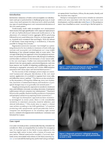
           tributing to angiogenic processes and cellular differentiation   Figure 1. Initial clinical photograph showing crown
                                                                discoloration and fracture on June 3, 2018.
           associated with tissue repair and dentinal development. 3-5
              For the success of this technique, several steps are consid-
           ered fundamental: adequate disinfection of the root canal
           system, application of a scaffold to organize host stem cells,
           and effective coronal sealing. 6-8  The literature also emphasiz-
           es the importance of long-term follow-up, since root matura-
           tion, formation of mineralized tissues, and resolution of peri-
           apical changes may occur gradually over several years. 1,9
              Despite promising results, regenerative endodontic treatment
           may yield different outcomes, including resolution of periapical
                                                  10
           radiolucency without complete root development.  Treatment
           failure is also possible, characterized by persistence of symptoms
           and periapical radiolucency, requiring further intervention. 11
              Although this therapeutic approach has been extensive-
           ly investigated in recent years, there is still considerable
           variability in treatment protocols, material selection, and
                            12
           clinical indications.  This study aims to report a clinical
           case, written according to the CARE guidelines,  of a patient
                                                13
           presenting with symptomatic apical periodontitis and in-
           complete root development in the maxillary right central
           incisor, treated with regenerative endodontic treatment and
           followed for 6 years.


           Case report

           A 12-year-old male patient, systemically healthy, attended the
           endodontic clinic accompanied by his mother for emergency
           care. He reported pain in the labial mucosa adjacent to the
           maxillary right central incisor, with a history of trauma one
           year earlier. Clinical examination revealed a fractured, dark-  Figure 2. Diagnostic periapical radiograph showing
                                                                incomplete root development and apical periodontitis
           ened crown associated with edema of the labial mucosa (Fig-  on June 3, 2018.
           ure 1). Cold sensitivity testing was performed using a refriger-

