


Page 49 - SPEMD_67-1
P. 49
rev port estomatol med dent cir maxilofac . 2026;67(1):40-46             43


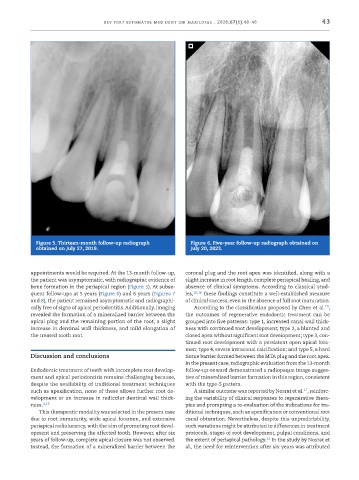
             Figure 5. Thirteen-month follow-up radiograph      Figure 6. Five-year follow-up radiograph obtained on
             obtained on July 27, 2019.                         July 20, 2023.


           appointments would be required. At the 13-month follow-up,   coronal plug and the root apex was identified, along with a
           the patient was asymptomatic, with radiographic evidence of   slight increase in root length, complete periapical healing, and
           bone formation in the periapical region (Figure 5). At subse-  absence of clinical symptoms. According to classical stud-
           quent follow-ups at 5 years (Figure 6) and 6 years (Figures 7   ies, 10,16  these findings constitute a well-established measure
           and 8), the patient remained asymptomatic and radiographi-  of clinical success, even in the absence of full root maturation.
                                                                                                             10
           cally free of signs of apical periodontitis. Additionally, imaging   According to the classification proposed by Chen et al. ,
           revealed the formation of a mineralized barrier between the   the outcomes of  regenerative endodontic treatment can be
           apical plug and the remaining portion of the root, a slight   grouped into five patterns: type 1, increased canal wall thick-
           increase in dentinal wall thickness, and mild elongation of   ness with continued root development; type 2, a blunted and
           the treated tooth root.                             closed apex without significant root development; type 3, con-
                                                               tinued root development with a persistent open apical fora-
                                                               men; type 4, severe intracanal calcification; and type 5, a hard
           Discussion and conclusions                          tissue barrier formed between the MTA plug and the root apex.
                                                               In the present case, radiographic evaluation from the 13-month
           Endodontic treatment of teeth with incomplete root develop-  follow-up onward demonstrated a radiopaque image sugges-
           ment and apical periodontitis remains challenging because,   tive of mineralized barrier formation in this region, consistent
           despite the availability of traditional treatment techniques   with the type-5 pattern.
                                                                                                      17
           such as apexification, none of these allows further root de-  A similar outcome was reported by Nosrat et al. , reinforc-
           velopment or an increase in radicular dentinal wall thick-  ing the variability of clinical responses to regenerative thera-
           ness. 1,15                                          pies and prompting a re-evaluation of the indications for tra-
              This therapeutic modality was selected in the present case   ditional techniques, such as apexification or conventional root
           due to root immaturity, wide apical foramen, and extensive   canal obturation. Nevertheless, despite this unpredictability,
           periapical radiolucency, with the aim of promoting root devel-  such variations might be attributed to differences in treatment
           opment and preserving the affected tooth. However, after six   protocols, stages of root development, pulpal conditions, and
           years of follow-up, complete apical closure was not observed.   the extent of periapical pathology.  In the study by Nosrat et
                                                                                         12
           Instead, the formation of a mineralized barrier between the   al., the need for reintervention after six years was attributed

