


Page 48 - SPEMD_67-1
P. 48
42                      rev port estomatol med dent cir maxilofac . 2026;67(1):40-46


                                       14
           dex (PAI) proposed by Ørstavik et al.  The patient had no sys-
           temic or hereditary diseases and no history of drug hypersen-
           sitivity. Regenerative endodontic treatment  was proposed.
             The patient and his legal guardian received detailed infor-
           mation about the proposed treatment and signed an informed
           consent form in accordance with the Brazilian National Health
           Council’s guidelines.
             Initially, local anesthesia was achieved using one cartridge
           of lidocaine hydrochloride with epinephrine 36 mg (Alpha-
           caine, DFL, Rio de Janeiro, Brazil), administered in the muco-
           buccal fold adjacent to the tooth and in the palatal gingival
           papilla. Access cavity preparation was performed with a spher-
           ical bur #1014 (KG Sorensen, Espírito Santo, Brazil), and cavity
           refinement was carried out with a non-cutting tapered bur
           #3083 (KG Sorensen, Espírito Santo, Brazil), under high-speed
           rotation with water cooling. Absolute isolation was achieved
           using a rubber dam sheet (Madeitex, São Paulo, Brazil) and
           clamp #211 (Golgran, São Paulo, Brazil).
             Chemical preparation was performed using 2.5% sodium
           hypochlorite solution (Asfer, São Paulo, Brazil), and no me-
           chanical instrumentation of the root canal was carried out.
           Prior to filling the canal with intracanal medication, drying
           was performed using sterile paper points size #80 (Dentsply
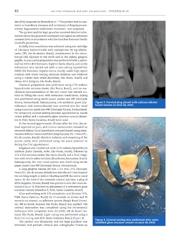
           Sirona, Switzerland). Subsequently, a bi-antibiotic paste (cip-  Figure 3. Cervical plug placed with calcium silicate-
           rofloxacin and metronidazole) was inserted into the canal   based cement on June 30, 2018.
           using a Lentulo spiral size #40 (Dentsply Sirona, Switzerland).
           For temporary coronal sealing between appointments, sterile
           cotton pellets and a restorative glass ionomer cement (Maxx-
           ion R, FGM, Santa Catarina, Brazil) were used.
             At the second appointment, 28 days after the first, the pa-
           tient reported no pain, and clinical examination revealed no
           intraoral edema. Local anesthesia was performed using mepi-
           vacaine without vasoconstrictor (Mepivacaine 3% – Nova DFL,
           Rio de Janeiro, Brazil). Absolute isolation and reopening of the
           access cavity were performed using the same protocol as
           during the first appointment.
             Irrigation was carried out with 2.5% sodium hypochlorite
           solution (Soda Clorada, Asfer, São Paulo, Brazil), followed by
           17% EDTA solution (Asfer, São Paulo, Brazil), and a final irriga-
           tion with sterile saline solution (Eurofarma, Amazonas, Brazil).
           Subsequently, the root canal system was dried using sterile
           paper points size #80 (Dentsply Sirona, Switzerland).
             A long gingival needle (38 mm × 0.4 mm, 27G; Dencojet,
           Nova DFL, Rio de Janeiro, Brazil) was introduced 2 mm beyond
           the working length to induce bleeding and fill the entire canal
           space. At the level of the cemento-enamel junction, a plug of
           MTA (Angelus, Paraná, Brazil) was placed to seal the canal en-
           trance (Figure 3), followed by placement of a restorative glass
           ionomer cement (Maxxion R, FGM, Santa Catarina, Brazil).
             After acid etching with 37% phosphoric acid (Condac 37%,
           FGM, Santa Catarina, Brazil) for 15 seconds on dentin and 30
           seconds on enamel, an adhesive system (Single Bond Univer-
           sal, 3M do Brasil, Sumaré, São Paulo, Brazil) was applied. The
           coronal restoration was completed using the incremental
           technique with composite resin A3 (Z350, 3M do Brasil, Su-
           maré, São Paulo, Brazil). Light curing was performed using a
           Radii Cal curing unit (SDI, Santa Catarina, Brazil) (Figure 4).  Figure 4. Coronal sealing was performed with resin-
             The patient was dismissed, and the legal guardian was   modified glass ionomer cement on June 30, 2018.
           informed that periodic clinical and radiographic follow-up

