


Page 50 - SPEMD_67-1
P. 50
44                      rev port estomatol med dent cir maxilofac . 2026;67(1):40-46


                                                                       18
                                                              Kahler et al.  emphasize that additional interventions are not
                                                              required when there is clear evidence of resolution of periapi-
                                                              cal pathology, even in the absence of significant root matura-
                                                              tion. This understanding supports the clinical management
                                                              adopted in the present report.
                                                                 The protocol described in this case followed the recom-
                                                              mendations of the  American  Association of Endodontists
                                                              (AAE) that were in effect at the time of treatment. 19,20  Ade-
                                                              quate disinfection of the root canal system is essential; how-
                                                              ever, conventional chemomechanical preparation may further
                                                                                                            21
                                                              weaken thin dentinal walls. Therefore, according to Wei et al. ,
                                                              minimal instrumentation may be indicated. Sodium hypochlo-
                                                              rite at low concentrations does not exert deleterious effects on
                                                              stem cells derived from the apical papilla, and its neutraliza-
                                                              tion before bleeding induction by a final rinse with sterile sa-
                                                              line solution is recommended. 22,23
                                                                 The use of 17% EDTA in regenerative endodontic proce-
                                                              dures plays an essential role by neutralizing sodium hypochlo-
                                                              rite and inducing the release of growth factors from dentin,
                                                              particularly transforming growth factor beta (TGF-β).  To fur-
                                                                                                       24
                                                              ther enhance canal disinfection, a bi-antibiotic paste was used
                                                              in this case, as it effectively inhibits bacterial growth within
                                                              the root canal system. 24
                                                                 Once a suitable environment for cellular activity has been
                                                              established, a scaffold must be introduced to enable interac-
                                                              tion between stem cells and growth factors. In the present
                                                              case, a blood clot was used as the scaffold, as it has demon-
                                                              strated satisfactory clinical outcomes, does not require addi-
                                                              tional equipment or resources, is easy to obtain, and presents
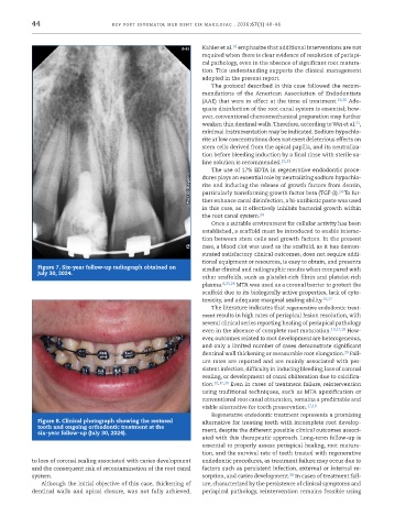
            Figure 7. Six-year follow-up radiograph obtained on   similar clinical and radiographic results when compared with
            July 30, 2024.
                                                              other scaffolds, such as platelet-rich fibrin and platelet-rich
                                                              plasma. 6,25,26  MTA was used as a coronal barrier to protect the
                                                              scaffold due to its biologically active properties, lack of cyto-
                                                              toxicity, and adequate marginal sealing ability. 16,27
                                                                 The literature indicates that regenerative endodontic treat-
                                                              ment results in high rates of periapical lesion resolution, with
                                                              several clinical series reporting healing of periapical pathology
                                                              even in the absence of complete root maturation. 10,17,18  How-
                                                              ever, outcomes related to root development are heterogeneous,
                                                              and only a limited number of cases demonstrate significant
                                                                                                         29
                                                              dentinal wall thickening or measurable root elongation.  Fail-
                                                              ure rates are reported and are mainly associated with per-
                                                              sistent infection, difficulty in inducing bleeding, loss of coronal
                                                              sealing, or development of canal obliteration due to calcifica-
                                                              tion. 10,17,30  Even in cases of treatment failure, reintervention
                                                              using traditional techniques, such as MTA apexification or
                                                              conventional root canal obturation, remains a predictable and
                                                              viable alternative for tooth preservation. 17,18
                                                                 Regenerative endodontic treatment represents a promising
            Figure 8. Clinical photograph showing the restored   alternative for treating teeth with incomplete root develop-
            tooth and ongoing orthodontic treatment at the
            six-year follow-up (July 30, 2024).               ment, despite the different possible clinical outcomes associ-
                                                              ated with this therapeutic approach. Long-term follow-up is
                                                              essential to properly assess periapical healing, root matura-
                                                              tion, and the survival rate of teeth treated with regenerative
           to loss of coronal sealing associated with caries development   endodontic procedures, as treatment failure may occur due to
           and the consequent risk of recontamination of the root canal   factors such as persistent infection, external or internal re-
                                                                                        28
           system.                                            sorption, and caries development.  In cases of treatment fail-
             Although the initial objective of this case, thickening of   ure, characterized by the persistence of clinical symptoms and
           dentinal  walls and  apical closure, was  not  fully achieved,   periapical pathology, reintervention remains feasible using

