


Page 42 - SPEMD_67-1
P. 42
36                      rev port estomatol med dent cir maxilofac . 2026;67(1):34-39


            Table 1. Differential diagnosis of a reddish exophytic gingival lesion
            Lesion            Key clinical features  Radiographic findings  Histopathology  Differentiation from Pgcg
            Pyogenic granuloma  Soft, bright red, lobulated,   No bone involvement  Granulation tissue with   More vascular, lacks giant
                              often ulcerated                         numerous capillaries  cells
            Peripheral ossifying   Firm pink or red nodule,   May show focal   Fibrous stroma with   Firm texture, presence of
            fibroma           interdental papilla  calcifications     mineralized tissue   calcifications
            Peripheral odontogenic   Slow-growing, firm lesion  No bone involvement  Fibrous tissue with   No multinucleated giant
            fibroma                                                   odontogenic rests    cells
            Hemangioma        Bluish-red, compressible,   No bone involvement  Proliferation of vascular   Positive blanching on
                              blanches on pressure                    channels             diascopy and possible blood
                                                                                           aspiration
            Central giant cell   Intraosseous origin  Radiolucency with cortical   Giant-cell–rich stroma  Originates within the bone
            granuloma                             thinning
            Brown tumor       Bone lesion associated with   Radiolucent lesion with bone   Giant-cell–rich lesion similar   Requires correlation with
            (hyperparathyroidism)  hyperparathyroidism; may   involvement  to central giant cell   systemic metabolic findings
                              mimic giant cell lesions                granuloma            (calcium, phosphate, ALP,
                                                                                           PTH)
           PGCG, peripheral giant cell granuloma.

                                                              affected area; the residual roots were extracted, and scaling
                                                              and root planning of adjacent teeth were performed. No de-
                                                              fective restorations or prosthetic factors were identified. Peri-
                                                              odontal evaluation revealed no significant tooth mobility or
                                                              deep periodontal pockets, and all adjacent teeth were pre-
                                                              served. This comprehensive approach aimed to eliminate po-
                                                              tential sources of chronic irritation and reduce the risk of re-
                                                              currence.
                                                                 Primary closure was achieved using interrupted sutures
                                                              with non-resorbable silk (3-0). The excised specimen was fixed


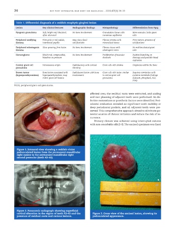
            Figure 1. Intraoral view showing a reddish-violet
            pedunculated lesion from the permanent mandibular
            right canine to the permanent mandibular right
            second premolar (teeth 43–45).


            Figure 2. Panoramic radiograph showing superficial
            cortical alteration in the region of teeth 43–45 and the   Figure 3. Gross view of the excised lesion, showing its
            presence of residual roots and carious lesions.     pedunculated appearance.

