
Page 12 - SPEMD_61-3
P. 12
102 rev port estomatol med dent cir maxilofac. 2020;61(3):97-105
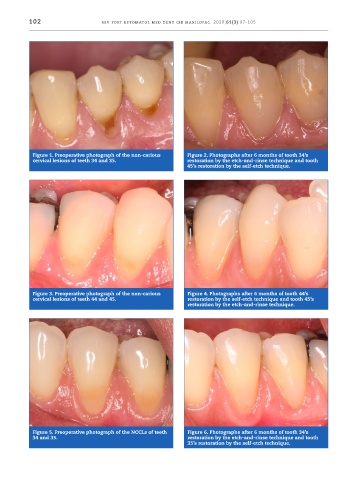
Figure 1. Preoperative photograph of the non -carious Figure 2. Photographs after 6 months of tooth 34’s
cervical lesions of teeth 34 and 35. restoration by the etch -and -rinse technique and tooth
45’s restoration by the self -etch technique.
Figure 3. Preoperative photograph of the non -carious Figure 4. Photographs after 6 months of tooth 44’s
cervical lesions of teeth 44 and 45. restoration by the self -etch technique and tooth 45’s
restoration by the etch -and -rinse technique.
Figure 5. Preoperative photograph of the NCCLs of teeth Figure 6. Photographs after 6 months of tooth 34’s
34 and 35. restoration by the etch -and -rinse technique and tooth
35’s restoration by the self -etch technique.