


Page 17 - SPEMD_62-3
P. 17
rev port estomatol med dent cir maxilofac . 2021;62(3):141-149         143


            Table 1. Inclusion and exclusion criteria
                              Inclusion Criteria                               Exclusion Criteria
            Be at least 18 years old                          Restored or non -vital anterior teeth
            Absence of smoking habits                         Presence of periodontal disease or caries lesions
            Willing to sign a consent form                    Being under orthodontic treatment
            Have at least one anterior tooth (maxillary or mandibular) equal or   Allergy to one of the components
            darker than A3,5 in VITA Classic shade guide measurable by a
            spectrophotometry                                 Pregnancy or lactating women
                                                              Anomalies of tooth development or presence of severe tooth
                                                              discoloration
                                                              History of any medical disease that may interfere with the study or
                                                              require special consideration
                                                              Poor oral hygiene


           brary of Medicine ClinicalTrials.gov website under the refer-  system (CIE L*a*b* values of the buccal tooth surface), and the
           ence number NCT03588871, in full compliance with the Hel-  difference (ΔE ) was calculated to determine bleaching effec-
                                                                         00
           sinki  World Medical  Association Declaration’s most recent   tiveness. Tooth whiteness was evaluated with a whiteness in-
           amendments. Patients were recruited consecutively, screened   dex (WI ) specifically established for dentistry and based on
                                                                     D
           according to inclusion and exclusion criteria (Table 1), signed   the CIELAB color notation system. The whiteness index was
           an informed consent form, and received professional dental   assessed before (WI ) and after (WI ) the bleaching treatment,
                                                                             D1
                                                                                          D2
           prophylaxis. Then, they were randomly allocated to one of   and then its difference was calculated (ΔWI ). 22
                                                                                                D
           two study groups (GraphPad QuickCals, http://www.graph-
           pad.com/quickcalcs/randomize1.cfm), according to different
           soft-tissue protection materials, for an in-office technique
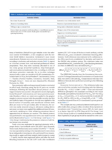
           protocol with a paint-on varnish at 6% HP concentration (Vi-  The CIEDE2000 formula, from the Commission Internatio-
                                         ®
           vaStyle Paint On PIus, lvoclarVivadent , Liechtenstein): Group   nale De l’Eclairage (International Commission on Illumination)
           1 – Vaseline (Purified Vaseline, Continente, SonaeMC, Maia,   (CIE), was used to calculate ΔE . Computations with this color
                                                                                      00
           Portugal); Group 2 – block-out resin (OpaldamTM, Ultradent   difference formula were performed according to the following
           Products, Inc, USA).                                equation: 23
              Two previously calibrated experienced dentists conducted   Parametric factors were set to 1. The whiteness index was
           in-office tooth bleaching using the 6% HP paint-on varnish   calculated before and after tooth bleaching with the following
           technique, following the described clinical protocol: an Op-  formula: WI  = 0.511L*-2.324a*-1.100b*.  Color and whiteness
                                                                                             24
                                                                        D
           tragate retractor was placed in the patient’s mouth (Optragate,   difference perception was assessed according to two major
                       ®
           lvoclarVivadent , Liechtenstein); soft-tissue protection mate-  thresholds: perceptibility threshold (PT for ΔE ; WPT for ΔWI )
                                                                                                 00
                                                                                                             D
           rials (Vaseline or block-out resin) were applied to the gingival   at ΔE  = 0.8 and ΔWI  = 0.72; and acceptability threshold (AT
                                                                               D
                                                                  00
           margin to prevent contact with the peroxide varnish; one thin   for ΔE ; WAT for ΔWI ) at ΔE  = 1.8 and ΔWI  = 2.60.  24-26  ΔWI
                                                                   00
                                                                                                 D
                                                                                    00
                                                                               D
                                                                                                              D
           and uniform layer of the paint-on varnish was applied to the   evaluation followed Perez et al.’s classification system present-
           buccal surface of maxillary and mandibular anterior teeth   ed in Table 2. 26
           (first pre-molar to first pre-molar); after 10 minutes, the var-  The sample size was previously determined based on our
           nish was easily removed using an ultrasonic scaling device;   pilot study’s data, using an online calculator (http://powerand-
           and paint-on layers were applied five additional times (10 min-  samplesize.com). 18  Considering a mean ΔWI for teeth equal
                                                                                                 D
           utes each), resulting in a total whitening procedure time of   to or darker than A3.5 in VITA Classical (9.9 for Group 1 and
                                          6
           approximately 60 minutes per session.  The clinical protocol   12.9 for Group 2) with a 3.2 standard deviation, we established
           was performed in two appointments with a one-week interval   that 20 patients (10 per group) would be needed for a two-sam-
           and is illustrated in Figures 1 to 6 for Group 1 and Figures 7 to
           14 for Group 2. Additionally, the presence or absence of soft-tis-               26
                                                                          D
           sue lesions was recorded.                            Table 2. ΔWI  classification system.
              Tooth color was evaluated by spectrophotometry with a                           ΔWID 50:50%
           proper device: SpectroShade micro (SS) (MHT Optic Research,   Visual grading     Threshold values
           Niederhasli, Switzerland; serial number HDL3973). 19-21   The de-  0: No difference   0.70
           vice was operated according to the manufacturer’s instructions
           by an independent investigator, who performed three measur-  1: Small difference      1.57
           ing rounds in upper and lower anterior teeth (canine to canine   2: Fairly acceptable difference  2.96
           – 12 teeth). Results were registered before and after the tooth   3: Hardly acceptable difference   5.69
           bleaching treatment in the CIE L*a*b* tooth color coordinates

