


Page 20 - SPEMD_62-3
P. 20
146                    rev port estomatol med dent cir maxilofac. 2021;62(3):141-149


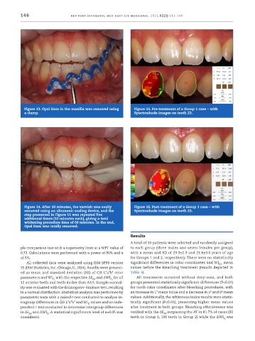
            Figure 13. Opal Dam in the maxilla was removed using   Figure 15. Pre ‑treatment of a Group 1 case – with
            a clamp.                                            Spectroshade images on tooth 23.


            Figure 14. After 10 minutes, the varnish was easily   Figure 16. Post ‑treatment of a Group 1 case – with
            removed using an ultrasonic scaling device, and the   Spectroshade images on tooth 23.
            step presented in Figure 11 was repeated five
            additional times (10 minutes each), giving a total
            whitening procedure time of 60 minutes. In the end,
            Opal Dam was totally removed. 6

                                                              Results
                                                              A total of 20 patients were selected and randomly assigned
           ple comparison test with a superiority limit at a WPT value of   to each group (three males and seven females per group),
           0.72. Calculations were performed with a power of 80% and α   with a mean and SD of 23.3±2.3 and 21.6±3.0 years of age
           of 5%.                                             for Groups 1 and 2, respectively. There were no statistically
             All collected data were analyzed using IBM SPSS version   significant differences in color coordinates and WI  mean
                                                                                                       D1
           25 (IBM Statistics, Inc. Chicago, IL, USA). Results were present-  values before the bleaching treatment (results depicted in
           ed as mean and standard deviation (SD) of CIE L*a*b* color   Table 3).
           parameters and WI  with the respective ΔE  and ΔWI  for all   All  treatments  occurred  without  drop-outs,  and  both
                          D,
                                            00
                                                     D,
           12 anterior teeth and teeth darker than A3.5. Sample normal-  groups presented statistically significant differences (P<0.01)
           ity was evaluated with the Kolmogorov-Smirnov test, resulting   for tooth color coordinates after bleaching procedures, with
           in a normal distribution. Statistical analysis was performed by   an increase in L* mean value and a decrease in a* and b* mean
           parametric tests with a paired t-test conducted to analyze in-  values. Additionally, the whiteness index results were statis-
           tragroup differences in CIE L*a*b* and WI  values and an inde-  tically significant (P<0.01), presenting higher mean values
                                          D
           pendent t-test conducted to determine intergroup differences   after treatment in both groups. Bleaching effectiveness was
           in ΔE  and ΔWI . A statistical significance level of α=0.05 was   verified with the ΔE surpassing the AT in 81.7% of cases (90
               00      D                                                      00
           considered.                                        teeth in Group 1; 106 teeth in Group 2) while the ΔWI was
                                                                                                         D

