
Page 35 - SPEMD_61-2
P. 35
rev port estomatol med dent cir maxilofac . 2020;61(2):72-78 73
Tratamento de malformação vascular labial com o laser de díodo
cirúrgico: Caso clínico
r e s u m o
Palavras-chave: O objetivo deste estudo é apresentar um caso clínico de hemangioma do lábio inferior tra-
Laser de díodo tado com sucesso, por laser de díodo. Paciente com 12 anos de idade com malformação
Excisão com laser vascular ocupando toda a zona vermelha do lábio inferior direito incluindo a face interna
Incisão com laser até à linha média. O tratamento cirúrgico foi efetuado em 2 sessões com o laser de díodo
Cirurgia a laser (GaAl) de 980 nm com fibra descartável (fiber tips) de Ø 400 µm. Houve reepitelização total
Hemangioma oral das feridas em aproximadamente 21 a 30 dias. Não ocorreu dor pós-operatória e o resultado
Cicatrização de feridas funcional e estético foi bom. O uso do laser de díodo nas anomalias vasculares orais pode
ser considerado uma boa opção de tratamento. (Rev Port Estomatol Med Dent Cir Maxilofac.
2020;61(2):72-78)
© 2020 Sociedade Portuguesa de Estomatologia e Medicina Dentária.
Published by SPEMD. This is an open access article under the CC BY-NC-ND license
(http://creativecommons.org/licenses/by-nc-nd/4.0/).
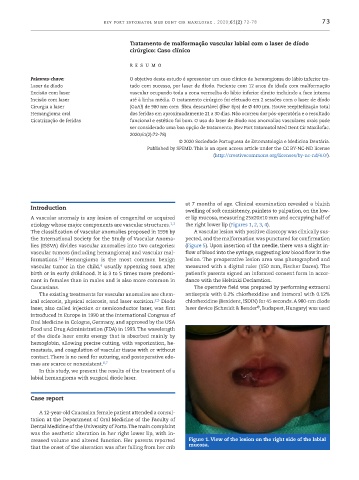
at 7 months of age. Clinical examination revealed a bluish
Introduction
swelling of soft consistency, painless to palpation, on the low-
A vascular anomaly is any lesion of congenital or acquired er lip mucosa, measuring 25x20x10 mm and occupying half of
etiology whose major components are vascular structures. 1,2 the right lower lip (Figures 1, 2, 3, 4).
The classification of vascular anomalies proposed in 1996 by A vascular lesion with positive diascopy was clinically sus-
the International Society for the Study of Vascular Anoma- pected, and the malformation was punctured for confirmation
lies (ISSVA) divides vascular anomalies into two categories: (Figure 5). Upon insertion of the needle, there was a slight in-
vascular tumors (including hemangioma) and vascular mal- flow of blood into the syringe, suggesting low blood flow in the
formations. 2,3 Hemangioma is the most common benign lesion. The preoperative lesion area was photographed and
4
vascular tumor in the child, usually appearing soon after measured with a digital ruler (150 mm, Fischer Darex). The
birth or in early childhood. It is 3 to 5 times more predomi- patient’s parents signed an informed consent form in accor-
nant in females than in males and is also more common in dance with the Helsinki Declaration.
Caucasians. The operative field was prepared by performing extraoral
The existing treatments for vascular anomalies are chem- antisepsis with 0.2% chlorhexidine and intraoral with 0.12%
ical sclerosis, physical sclerosis, and laser excision. 2,5 Diode chlorhexidine (Bexident, ISDIN) for 45 seconds. A 980 -nm diode
®
laser, also called injection or semiconductor laser, was first laser device (Schmidt & Bender , Budapest, Hungary) was used
introduced in Europe in 1990 at the International Congress of
Oral Medicine in Cologne, Germany, and approved by the USA
Food and Drug Administration (FDA) in 1993. The wavelength
of the diode laser emits energy that is absorbed mainly by
hemoglobin, allowing precise cutting, with vaporization, he-
mostasis, and coagulation of vascular tissue with or without
contact. There is no need for suturing, and postoperative ede-
mas are scarce or nonexistent. 6,7
In this study, we present the results of the treatment of a
labial hemangioma with surgical diode laser.
Case report
A 12 -year -old Caucasian female patient attended a consul-
tation at the Department of Oral Medicine of the Faculty of
Dental Medicine of the University of Porto. The main complaint
was the aesthetic alteration in her right lower lip, with in-
creased volume and altered function. Her parents reported Figure 1. View of the lesion on the right side of the labial
that the onset of the alteration was after falling from her crib mucosa.