
Page 38 - SPEMD_61-2
P. 38
76 rev port estomatol med dent cir maxilofac. 2020;61(2):72-78
The result confirmed the clinical diagnosis of vascular heman-
gioma (Figures 10, 11). The wound was left open for healing by
second intention, without the use of any surgical cement. Post-
operative care included the application of a 0.2% chlorhexidine
gel (Bexident Post Gel, ISDIN) on the wound.
In the second session of the treatment, we approached
the red zone of the lip, using the same parameters as in the
first session (Figure 12). An SPF 50 cream (Avéne) was applied
to the labial mucosa sensitive areas for 21 days to 3 months
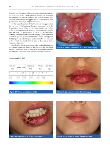
after surgery. The patient was followed up 30 days post-
-surgery. In the first three days after surgery, a pain evaluation
scale was applied, standardized with a visual analog scale
(VAS) (Figure 13). Following the instructions for use provided
to the patient, she had to draw a cross at the point of maxi-
mum pain intensity.
Shortly after the surgery, a scorched area of carbonized and
edematous tissue was observed. By the third day, no visible Figure 15. Wound appearance at 11 days post ‑surgery.
areas of carbonized tissue were detected. The lesions were well
delimited with some necrotic material covering the wound. It
Figure 13. Visual Analog Scale (VAS). Figure 16. Lip aspect at 1 month post ‑surgery.
Figure 14. Appearance at 3 days post ‑surgery. Figure 17. Lip aspect at 3 months post ‑surgery.