
Page 37 - SPEMD_61-2
P. 37
rev port estomatol med dent cir maxilofac . 2020;61(2):72-78 75
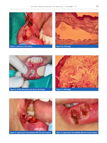
Figure 7. Exertion of the lesion. Figure 10. Histology.
Figure 8. Visible bleaching and tissue shrinkage. Figure 11. Histology.
Figure 9. Appearance immediately after the first surgery. Figure 12. Appearance immediately after the second surgery.