
Page 36 - SPEMD_61-2
P. 36
74 rev port estomatol med dent cir maxilofac. 2020;61(2):72-78
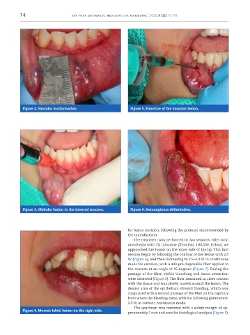
Figure 2. Vascular malformation. Figure 5. Puncture of the vascular lesion.
Figure 3. Globular lesion in the internal mucosa. Figure 6. Hemangioma delimitation.
for lesion excision, following the protocol recommended by
the manufacturer.
The treatment was performed in two sessions. After local
anesthesia with 2% lidocaine (Xilonibsa 1:80,000, Inibsa), we
approached the tumor on the inner side of the lip. This first
session began by following the contour of the lesion with 2.0
W (Figure 6), and then increasing to 2.5–3.0 W in continuous
mode for excision, with a 400 -μm disposable fiber applied to
the mucosa at an angle of 45 degrees (Figure 7). During the
passage of the fiber, visible bleaching and tissue retraction
were observed (Figure 8). The fiber remained in close contact
with the tissue and was slowly moved around the lesion. The
deeper area of the epithelium showed bleeding, which was
coagulated with a second passage of the fiber on the capillary
from where the bleeding came, with the following parameters:
2.0 W, in contact, continuous mode.
The specimen was removed with a safety margin of ap-
Figure 4. Mucosa labial lesion on the right side.
proximately 1 mm and sent for histological analysis (Figure 9).