
Page 50 - SPEMD_59-4
P. 50
222 rev port estomatol med dent cir maxilofac. 2018;59(4):221-224
ção anticonvulsivante. Diretrizes sobre hábitos de higiene oral e alimentar, bem como
aplicação tópica de verniz fluoretado foram implementadas. A hiperplasia foi revertida
em 2 semanas e o paciente tem recebido acompanhamento. O paciente encontra-se em
monitorização e houve melhoria de sua condição oral. (Rev Port Estomatol Med Dent Cir
Maxilofac. 2018;59(4):221-224)
© 2018 Sociedade Portuguesa de Estomatologia e Medicina Dentária.
Published by SPEMD. This is an open access article under the CC BY-NC-ND license
(http://creativecommons.org/licenses/by-nc-nd/4.0/).
had been born with hydrocephalus, facial changes, and cleft
Introduction
lip/palate.
Peters-plus syndrome, also known as Krause-Kivlin syn- The patient had already undergone 10 surgical interven-
drome, is a rare congenital disorder of glycosylation with an tions, including cheiloplasty to correct the cleft lip at the age
1,2
autosomal recessive pattern. Its exact incidence is still un- of 6 months, two interventions for the implantation of ventric-
known, and fewer than 75 cases with this anomaly have been uloperitoneal shunt valves to relieve intracranial pressure, two
identified in the medical literature. 3 to correct an inguinal hernia, two surgeries for eyelid recon-
Peters-plus syndrome can be diagnosed clinically by the struction, and three external and middle ear surgeries.
presence of Peters anomaly – a congenital corneal opacity sec- The patient had undergone cardiac examinations for the
ondary to a defect in neural crest cell migration that causes a diagnosis of possible heart abnormalities, which are common-
malformation of the anterior eye segment, associated with ly associated with the syndrome, but no abnormalities were
other symptoms. These symptoms include a delayed psycho- found. There was no report of food or medication hypersensi-
motor development, variable degrees of mental retardation, tivity. The anticonvulsant drug Gardenal (40mg/mL) had been
®
disproportionate short stature, and some facial features such continuously administered orally twice a day over the previous
as cleft lip or palate (present in about half of the cases), hyper- 10 months, together with vitamin C. At the extraoral clinical
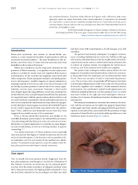
telorism, narrow eyes, prominent forehead, a thin/cupid’s examination, the syndrome’s typical facial appearance was
bow-shaped upper lip, long philtrum, small ears, hearing loss, observed, including bilateral corneal opacity (Figure 1), which
broad/webbed neck, and joint hyperextensibility. The presence was more visible in the right eye with consequent vision re-
of congenital heart defects, genitourinary abnormalities such duction, short stature, delayed psychomotor development and
as cryptorchidism, hypoplastic clitoris and hydronephrosis, as hearing loss.
well as structural brain malformations may affect the progno- The intraoral examination revealed the presence of unilat-
sis of individuals. Homozygous mutations of the B3GALTL gene eral cleft lip and palate on the right side, gingival hyperplasia
in the 13q12.3 region have been associated with this pheno- in the upper and lower arches, and gingival bleeding on brush-
4
type. Toxic, infectious and traumatic events have also been ing due to phenobarbital, associated with the presence of large
suggested as possible causes of this syndrome. 5 amounts of dental biofilm, and active white spot lesions in the
Little is known about this syndrome and studies in the deciduous upper incisors (Figures 2 and 3).
scientific literature, particularly in the dental field, are scarce.
Thus, knowing and identifying its main clinical and oral man-
ifestations is of great importance to establish an early diagno-
sis and rehabilitation, as well as referring families to genetic
counseling. 2,5-6
The dental treatment of these patients must especially
take into account the degree of mental retardation, the clinical
and oral manifestations of the disease, such as possible cardi-
ac anomalies, and the oral impact of the medications admin-
istered. The present study reports a case of a patient with Pe-
ters-plus syndrome submitted to dental treatment.
Case report
The 15-month-old male patient, B.S.M, diagnosed with Pe-
ters-plus syndrome, was brought to the School of Dentistry of
the Federal University of Amazonas by his paternal grand-
mother, with the main complaint of ‘gingival swelling’ since
birth. During anamnesis, the legal guardian informed us that Figure 1. Ocular changes, such as corneal opacity.
she had been referred by the neurologist and that the child