
Page 51 - SPEMD_59-4
P. 51
rev port estomatol med dent cir maxilofac . 2018;59(4):221-224 223
5
mutations in the B3GALTL gene, located at chromosome 13.
Therefore, the patient was under genetic follow-up to investi-
gate the sequential analysis of the gene and the molecular
diagnosis of a possible mutation; when the latter is not iden-
tified, deletion or duplication should be investigated. 3,6
On the other hand, studies have reported cases of Pe-
ters-plus syndrome with characteristic clinical manifestations
and lack of mutation of the B3GALTL gene. 4,13 However, in
these cases, not all the typical characteristics of the syndrome
are observed. The importance of the molecular diagnosis of
possible mutations is related to genetic counseling for future
pregnancies. Uncovering its causes, which are still unclear,
3
represents a potential chance for enhanced treatment and
prevention through genetic counseling or possible preventive
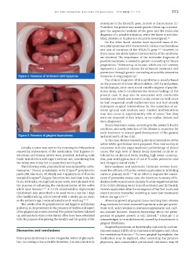
Figure 2. Presence of unilateral cleft lip/palate. measures during pregnancy. 5
The clinical diagnosis of this syndrome is usually based
on the presence of ocular abnormalities, cleft lip and palate,
facial changes, short stature and variable degrees of psycho-
motor delay, which corroborate the clinical findings of the
present case. It may also be associated with conductive
hearing loss, which was present in this patient as well, since
he had congenital small malformed ears and had already
undergone surgical interventions for the correction of ex-
ternal (pinna) and medium ears. Cardiac malformations
3
may also occur in approximately 33% of cases, but they
were not observed in this infant, as no cardiac defects had
been diagnosed.
Dental treatment varies according to the patient’s health
condition, and early detection of the disease is important for
early treatment to ensure good development of the general
Figure 3. Presence of gingival hyperplasia. and oral health of the child.
In this case, dietary counseling and remineralization of the
active white spot lesions were proposed. This relationship is
Initially, a letter was sent to the neurologist of the patient consistent with the sugar-mediated pathobiology of dental
requesting replacement of the medication. Oral hygiene in- caries. The high level of dental decay detected could be at-
structions and dietary advice were provided, including solid tributed to on-demand bottle feeding, high sweet consump-
foods’ introduction and sugar’s rational use, considering that tion, poor oral hygiene, lack of use of fluoride prevention and
the infant was solely fed on pasty food and liquids. lack of regular dental visits. 14
The following week, phenobarbital was replaced by carba- Meta-analyses and systematic literature reviews docu-
®
7
mazepine. Dental prophylaxis with Clinpro prophylactic ment the efficacy of fluoride varnish application in inhibiting
paste (3M, São Paulo, SP, Brazil) and 4 applications of fluoride caries in primary teeth. 8-10 In an effort to improve the consis-
®
varnish (Duraphat , Colgate-Palmolive Ind. And Com. Ltda, São tency of preventive dental care, the American Academy of Pe-
Paulo, SP, Brazil), one application per week, were planned with diatrics (AAP) recommends: (a) daily fluoride supplementation
the purpose of enhancing the remineralization of the active if the child’s drinking water is not fluoridated; and (b) fluoride
white spot lesions. 8-10 A 0.12% chlorhexidine digluconate varnish application after the emergence of the first tooth and
mouthwash was prescribed to be used twice a day for 7 days every 6 months thereafter, receiving at least four treatments
after toothbrushing, administered with a sterile gauze swab, before the age of 4. 8,15
as the patient’s age contraindicated mouth washing. 11-12 Abnormal growth of gingival tissue resulting from adverse
Two weeks after drug replacement and hygiene and dietary drug reactions has been observed in patients undergoing treat-
guidance, an improvement in the presence of biofilm and aspect ment with anticonvulsants, immunosuppressants and calci-
of the gingival tissue were observed. The patient has been followed um channel blockers. However, knowledge about the patho-
up, and quarterly visits to the dental office have been scheduled genesis of gingival growth is still limited, although it is
16
with the purpose of improving the family’s and his quality of life. acknowledged to be multifactorial, caused by disturbances in
gingival fibroblasts.
Gingival hyperplasia or hypertrophy induced by anticon-
Discussion and conclusions vulsants makes it difficult to maintain oral hygiene and, often,
17
the masticatory function. To treat gingival hyperplasia, the
Peters-plus syndrome is a rare congenital defect of glycosyla- medication may be replaced, after consulting the patient’s
tion. According to the scientific literature, it is associated with physician, and conservative periodontal treatment may be