
Page 58 - SPEMD_59-2
P. 58
116 rev port estomatol med dent cir maxilofac. 2018;59(2):115-118
rido para queratoacantoma gigante. O objetivo deste estudo é analisar as características
clinicopatológicas do queratoacantoma gigante de cabeça e pescoço, revisando os estudos
publicados na literatura inglesa e relatando um novo caso da lesão que apareceu no lábio.
(Rev Port Estomatol Med Dent Cir Maxilofac. 2018;59(2):115-118)
© 2017 Sociedade Portuguesa de Estomatologia e Medicina Dentária.
Publicado por SPEMD. Este é um artigo Open Access sob uma licença CC BY-NC-ND
(http://creativecommons.org/licenses/by-nc-nd/4.0/).
Introduction Case report
Keratoacanthoma (KA) is a rapid growth tumor character- A 64-year-old male patient was referred to the oral pathology
ized by its composition of well-differentiated keratinizing service of the João de Barros Barreto University Hospital, Belém,
squamous cells, which originate from the pilosebaceous Brazil, chiefly complaining of a 1-month rapidly growing swell-
follicles, and its etiology is not identified. 1-4 It is commonly ing on his lower lip. The patient had a 1-year history of a lesion
presented as a self-limiting lesion and has strong clinical in the lower lip when, in the previous 4 weeks, he noticed a
and histopathological similarities with squamous cell car- rapidly growing swelling and sought specialized assistance.
cinoma. 2,3,6
Head and neck KAs are common and account for around
1,3
70% of all KAs. Classically, the lesion is presented as a dome-
6
shaped nodule with an ulcerated crater in its center. Giant KA
is a rare variant of KA with a size exceeding 2 cm. 1,7 It has a
8
main predilection for the nose and eyelid as primary sites,
occurs more commonly in men, and most of the lesions are
observed in sun-exposed skin. 1,4,6
Owing to the rarity of the head and neck giant KA, few
case reports and clinical series have been published de-
scribing its biological behavior on the head and neck region.
The aim of this work is to analyze the clinicopathological
characteristics of the head and neck keratoacanthoma by
reviewing all the case reports and clinical series previously
published in English and reporting a rare new case in the
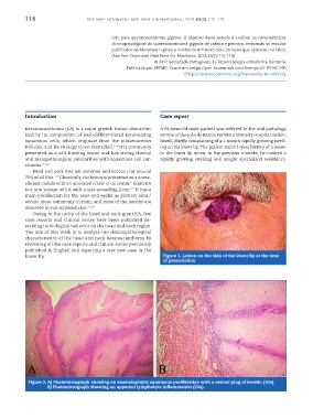
lower lip. Figure 1. Lesion on the skin of the lower lip at the time
of presentation
Figure 2. A) Photomicrograph showing an exoendophytic squamous proliferation with a central plug of keratin (10x).
B) Photomicrograph showing an apparent lymphocyte inflammation (20x).