
Page 59 - SPEMD_59-2
P. 59
rev port estomatol med dent cir maxilofac . 2018;59(2):115-118 117
rently presents a mean size of 3.93 cm in diameter. Patients
are usually in the sixth and seventh decade of life, with a mean
age of 59.1 years old, and are mainly males. 1,4,6,12,13 Studies
have reported that those tumors have a predilection for cos-
metically sensitive areas, including the nose and the eye-
lids. 8,14 Based on our literature analysis, the nose, the cheek
and the lower lip are the locations most commonly affected
by the head and neck giant KA.
The histopathological diagnosis of KA is further compli-
cated by its histological similarities with the squamous cell
carcinoma. 2,3,5 Histopathological features vary with the stage
3
of evolution. An early lesion is reported to increase rapidly,
generating cell differentiation with a high proliferation index.
The analysis varies according to the atypical mitosis, loss of
polarity, individual cell keratinization and hyperchromatic
cells of the specimen. 2,3,11 It is possibly associated with the
Figure 3. Tumor resection after surgical excision time of evolution. The KA is a lesion characterized by a rapid
and progressive growth. 4,6,11,15 Our analysis revealed a mean
evolution time of 15.1 months, although 73.3% of the cases
The patient had no history of trauma, infection, tumors or pri- presented a rapid progression in less than 3 days. Thus, while
or radiation. His medical history was noncontributory. tumor progression occurs, the degree of cell proliferation in-
Physical examination showed no abnormalities. No lymph creases and, consequently, it becomes increasingly similar to
node infarction was observed in the head and neck after palpa- the squamous cell carcinoma. 2,7 In the case report here pre-
tion. Clinically, the lesion exhibited an exophytic and peduncu- sented, the surgical specimen was reviewed carefully, confirm-
lated nodular appearance, with a verrucous surface and a violet ing the diagnosis of giant KA.
coloration, measuring 3.2 x 2.5 in diameter (Figure 1). No lymph Numerous treatment methods have been used in patients
nodes were observed. The patient did not refer pain in the area, with giant KA. Due to the tendency of KA to spontaneously re-
and the surrounding mucosa had a regular appearance. gress, the efficiency of some treatments is difficult to evalu-
An incisional biopsy was performed under local anesthesia, ate. 5,7,12,14,16 Only 12.5% of the patients were followed solely by
and the histopathological exam revealed tumor islands with en- observation, and all of them presented total tumor regression
larged keratinocytes formed by a downgrowth of squamous epi- after long follow -up times. When no type of treatment is em-
thelium with an increased amount of central keratin. The epider- ployed, there is a risk of the lesion invading adjacent tissues and
2,7
mis protruded irregularly and deeply into the dermis. Lymphocyte aesthetic commitment. The non -surgical treatments mainly
infiltration in the dermis was also observed (Figure 2). The sug- used for the regression of the lesion were the intralesional in-
17
gestive diagnosis of KA was made due to the histopathological terferon alpha 2a and methotrexate. 6,18 Taking into account
features. Then, the lesion was excised with a 1-mm margin, and the aggressive biological behavior of the head and neck giant
the histopathological exam of the whole lesion led to the diag- KA, surgical excision was the most used treatment approach
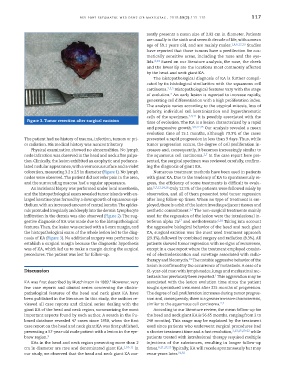
nosis of KA (Figure 3). No additional surgery was performed to (29.1%), followed by combined surgery and radiation (8.3%). All
establish a surgical margin because the diagnostic hypothesis patients showed tumor regression with no signs of recurrence,
was of KA, which led us to make a margin during the surgical except in a case report where the treatment employed consist-
procedures. The patient was lost for follow-up. ed of electrodesiccation and curettage associated with radio-
therapy and bleomycin. The notable aggressive behavior of the
19
lesion is confirmed by the occurrence of metastasis. A case of a
Discussion 61 -year -old man with lymph nodes, lungs and mediastinal me-
2
tastasis has previously been reported. This aggravation may be
8
KA was first described by Hutchinson in 1889. However, very associated with the lesion evolution time since the patient
few case reports and clinical series concerning the clinico- sought specialized treatment after 325 months of progression.
pathological features of the head and neck giant KA have The degree of cell proliferation increases during tumor progres-
been published in the literature. In this study, the authors re- sion and, consequently, there is a greater invasive characteristic,
viewed all case reports and clinical series dealing with the similar to the squamous cell carcinoma. 2,7
giant KA of the head and neck region, summarizing the most According to our literature review, the mean follow -up for
important aspects found by each author. A search in the Pu- the head and neck giant KA is 56.65 months, ranging from 2 to
bmed database revealed 47 cases since 1958, when the first 240 months). This range may be explained by the treatment
case report on the head and neck giant KA was first published, used since patients who underwent surgical procedures had
presenting a 57-year-old male patient with a lesion in the eye- a shorter treatment time and a fast resolution, 3,13,15,20-22 while
brow region. 9 patients treated with intralesional therapy required multiple
KAs in the head and neck region presenting more than 2 injections of the substances, resulting in longer follow -up
cm in diameter are rare and denominated giant KA. 1,10-11 In times. 6,11,17,23 Typically, KA will recede spontaneously but may
our study, we observed that the head and neck giant KA cur- recur years later. 31,32