
Page 54 - SPEMD_59-2
P. 54
112 rev port estomatol med dent cir maxilofac. 2018;59(2):107-114
Contrary to what occurs with other teeth, and as proven by
the clinical cases here presented, eruptive disorders involving the
3
MCI are frequently detected at the early mixed dentition phase.
Detailed clinical history and both clinical and radiographic ex-
2
aminations are pivotal for making a correct diagnosis. In partic-
ular, the clinical history is invaluable to screen for possible local
or systemic pathologies or the occurrence of trauma during child-
hood. 10,13 In Case 2, the patient’s history prior to tooth impaction,
in association with a traumatic episode involving the MCI’s pre-
decessor, demonstrates that traumatology is an important etio-
logical factor of this pathology and is usually responsible for the
dilaceration of MCIs that prompts impaction. 14,15 Dilaceration
refers to the displacement of a tooth’s root in relation to its nor-
16
mal alignment with the crown. Such deformity can pose com-
plications for impacted tooth treatment since the curved root can
affect the adjacent teeth or enter the labial cortical bone, which
causes pulp and periapical problems. 17,18 Case 2 presented an
MCI with an apical dilaceration of moderate severity, and its
treatment was achieved without complications, probably be-
cause during traction the apex slipped into the bone marrow,
despite being close to the intermaxillary suture.
When impaction of an MCI is suspected, it is essential to
search for certain indications of impaction in clinical exam-
inations – namely, an asymmetrical eruption with the homol-
ogous contralateral for roughly 6 months, the alteration of the
sequence or chronology of eruption, deciduous tooth reten-
tion, midline deviation, loss of space, and elevations in the soft
palatine tissue or labial mucosa. Following a careful clinical
13
examination, it is imperative to use a complementary means
of diagnosis. In all clinical cases presented, panoramic radiog-
raphy and lateral teleradiography were important methods for
studying the impacted MCI and general orthodontics.
With all the requisite information, it is possible to establish
a diagnosis and elaborate a treatment plan. Once complete,
the symbiosis of several medical specialties may be neces-
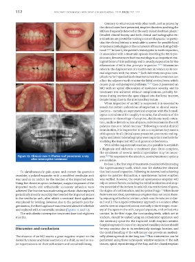
Figure 12. Clinical case 3: Photos and panoramic x-ray sary. 19,20 In response to the situation, several treatment options
after interceptive treatment
are available.
In Case 1, the first step of treatment consisted of eliminating
the supernumerary tooth, which was the obstructive element
To simultaneously gain space and correct the posterior that had caused impaction. Following its removal and achieving
crossbite, a palatal expander with a modified vestibular arch space via palatine disjunction, a spontaneous incisor eruption
was used as an anchor for the traction of the impacted tooth. was verified. However, the eventual spontaneous eruption will
Using the closed eruption technique, surgical exposure of the rely on several factors, including the initial localization and erup-
impacted tooth and orthodontic accessory adhesion were tive potential of the incisor, its axial tilt, the restrictions of space,
21
achieved. The traction was made using an elastic chain replaced the degree of root formation, and the patient’s age. When those
periodically (mostly monthly) that bonded the impacted incisor factors are not ideal, spontaneous eruption does not occur, there-
to the vestibular arch, after which a sectional fixed appliance by requiring orthodontic traction, which was the solution in Cas-
was placed for leveling. However, due to the patient’s poor hy- es 2 and 3. The surgical orthodontic approach is a solution often
gienization, the fixed appliance was removed ahead of schedule used to save an impacted incisor, normally in three stages: recov-
and replaced with a removable retainer (Figures 11 and 12). ery of the space in the arch, surgical exposure, and orthodontic
The orthodontic interceptive treatment time took eighteen traction. In the first stage, the surrounding teeth, which act as
months. anchors, should be united using an orthodontic appliance and
the necessary space for the impacted tooth eruption created.
Surgical exposure in case of an impacted maxillary incisor should
Discussion and conclusions be very cautious due to its aesthetically strategic location, and
the careful handling of the soft tissues can provide an aestheti-
22
The absence of an MCI exerts a great negative impact on the cally pleasing result in the long run. That intervention can be
dental functions and facial aesthetics of a child, as well as ma- performed using three techniques: window excision of the soft
jor repercussions on their self-esteem and social well-being. tissues, apical repositioning of the flap, and the closed eruption