
Page 50 - SPEMD_59-2
P. 50
108 rev port estomatol med dent cir maxilofac. 2018;59(2):107-114
nam-se imperativos. Uma das principais abordagens passa pela exposição cirúrgica, aber-
tura ortodôntica de espaço e tração do incisivo para a sua posição normal, o que acarreta
benefícios como a manutenção do dente e a possibilidade de manutenção do osso alveolar.
Este artigo apresenta a análise de três casos clínicos de indivíduos portadoras de um inci-
sivo central maxilar impactado com causa de impactação distinta nomeadamente dente
supranumerário, dilaceração da raiz e desvio de erupção, submetidos a tratamento e desta-
cando a tração ortodôntica. (Rev Port Estomatol Med Dent Cir Maxilofac. 2018;59(2):107-114)
© 2017 Sociedade Portuguesa de Estomatologia e Medicina Dentária.
Publicado por SPEMD. Este é um artigo Open Access sob uma licença CC BY-NC-ND
(http://creativecommons.org/licenses/by-nc-nd/4.0/).
Introduction Case report
The maxillary incisors definitively shape a person’s smile, Clinical Case 1
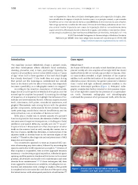
and their development affects children’s facial aesthetics, An 8-year-old female at an early mixed dentition phase com-
1
chewing, phonetics, and, in turn, psychology. Typically, the plained mainly of a late eruption of her right MCI. Her dental
eruption of a maxillary central incisor (MCI) occurs at 7 years medical history did not include any accident or trauma. Clini-
2
of age, when half to three-quarters of the root’s final length cal examination revealed a slight deviation of the superior
has already developed. If the tooth does not erupt during midline tooth and the inclination of the adjacent teeth to the
3
that period and the homologous contralateral has already edentulous space. Moreover, the patient presented a bilateral
been in the dental arch for about 6 months, then impaction class I molar and right unilateral posterior crossbite in the
4
becomes a possibility, as a radiographic control can suggest. maximum intercuspal position (Figure 1). Periapical radio-
According to the American Association of Orthodontists, graphic examination further revealed an intraosseous impac-
impaction is a tooth’s partial or total lack of eruption once the tion of her right MCI caused by the presence of a supernumer-
normal age for eruption has passed. Concerning the etiology ary tooth. Panoramic radiography and teleradiography
5
of impaction, it is important to highlight the influence of he- confirmed the presence of all permanent teeth, with the im-
reditary and environmental factors. Whereas supernumerary
teeth, odontomas, cleft palate, craniofacial syndromes, and
gingival fibromatosis rank among factors with the greatest
genetic components, environmental factors include trauma,
cystic pathology, maxillofacial skeletal insufficiency, and the
6
early extraction, loss, or retention of deciduous teeth.
MCIs play a crucial role in several aspects of a person’s
functioning and, for that reason, the absence of either of them
can pose several negative consequences, including poor facial
7
aesthetics and speech difficulty, namely with the “s” sound.
Eruption failure in those teeth can affect the eruption of other
teeth on the anterior level as well, namely the canine, due to
the loss of space, middle line deviation, or reabsorption of the
adjacent tooth or alveolar bone in the anterior region of the
maxilla. Therefore, the diagnosis and early treatment of MCI
8,9
impaction are imperative.
The conservative approach to treat an impacted MCI con-
sists of extracting any obstruction, followed by recovering the
10
space to enable the tooth’s spontaneous eruption. However, if
that eruption is impossible, then another approach becomes
necessary – one involving surgical exposition, orthodontic space
opening, and the posterior traction of the incisor to its normal
position, all of which can benefit tooth maintenance and even
alveolar bone maintenance. 11,12 If those techniques fail, then
more aggressive treatments are available, including incisor ex-
traction and restoration with a bridge or implant after growth
is completed or incisor extraction and closure of the space using Figure 1. Clinical case 1: Photos before treatment;
crossbite and right upper incisor inclusion
the lateral incisor with subsequent prosthetic rehabilitation. (11)