
Page 52 - SPEMD_59-2
P. 52
110 rev port estomatol med dent cir maxilofac. 2018;59(2):107-114
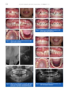
Figure 7. Clinical case 2: Orthodontic treatment with
partial upper fixed appliance
Figure 5. Clinical case 2: Photos before treatment; right
upper incisor inclusion
Figure 6. Clinical case 2: Periapical x-ray with right upper Figure 8. Clinical case 2: Photos and panoramic x-ray
incisor root dilaceration; teleradiography and after interceptive treatment
panoramic x-ray before orthodontic treatment