
Page 51 - SPEMD_59-2
P. 51
rev port estomatol med dent cir maxilofac . 2018;59(2):107-114 109
Figure 2. Clinical case 1: Periapical x-ray with a
mesiodens; teleradiography and panoramic
x-ray after mesiodens extraction
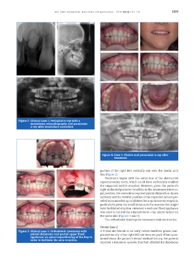
Figure 4. Case 1: Photos and panoramic x-ray after
treatment
paction of the right MCI vertically and over the dental arch
line (Figure 2).
Treatment began with the extraction of the obstructive
supernumerary tooth, which could have sufficiently enabled
the impacted tooth’s eruption. However, given the patient’s
right unilateral posterior crossbite in the maximum intercus-
pal position, the correction required palatal disjunction. Space
recovery and the vertical position of the impacted incisor pro-
vided accommodating conditions for a spontaneous eruption,
particularly given the small incision in the mucosa that might
have facilitated eruption. However, a sectional fixed appliance
was used to control the adjacent tooth—the lateral incisor on
the same side (Figures 3 and 4).
The orthodontic interceptive treatment took six months.
Clinical Case 2
Figure 3. Clinical case 1: Orthodontic treatment with A 9-year-old female at an early mixed dentition phase com-
palatal disjunctor and partial upper fixed plained mainly of her right MCI not have erupted. When ques-
appliance; an apical repositioning of the flap in tioned about the patient’s dental medical history, her parents
order to facilitate the auto-eruption.
reported a traumatic episode that had affected the deciduous