
Page 53 - SPEMD_59-2
P. 53
rev port estomatol med dent cir maxilofac . 2018;59(2):107-114 111
incisors. Clinical and radiographical examination confirmed
the right MCI’s dilaceration and impaction. The patient exhib-
ited a deviation of the upper dental line to the right consider-
ing the lower dental midline, a class I molar, and space in the
dental arch for the incisor’s eruption (Figure 5). Panoramic ra-
diography and lateral teleradiography revealed the impacted
right MCI with the evident root dilaceration (Figure 6).
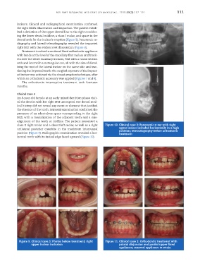
Treatment involved a sectional fixed orthodontic appliance
with bands at the level of the maxillary first molars and brack-
ets over the other maxillary incisors, first with a round section
arch and later with a rectangular one, all with the aim of distal-
izing the root of the lateral incisor on the same side and trac-
tioning the impacted tooth. The surgical exposure of the impact-
ed incisor was achieved via the closed eruption technique, after
which an orthodontic accessory was applied (Figures 7 and 8).
The orthodontic interceptive treatment took fourteen
months.
Clinical Case 3
An 8-year-old female at an early mixed dentition phase visit-
ed the dentist with her right MCI unerupted. Her dental med-
ical history did not reveal any event or element that justified
the absence of the tooth. Intraoral examination confirmed the
presence of an edentulous space corresponding to the right
MCI, with a mesialization of the adjacent teeth and a mis-
alignment of the teeth at midline. The patient presented a
class II right molar and a class I left molar, as well as a right Figure 10. Clinical case 3: Panoramic x-ray with right
unilateral posterior crossbite in the maximum intercuspal upper incisor included horizontally in a high
position; teleradiography before orthodontic
position (Figure 9). Radiographic examination revealed a hor- treatment
izontal tooth with its incisal edge faced upward (Figure 10).
Figure 9. Clinical case 3: Photos before treatment; right Figure 11. Clinical case 3: Orthodontic treatment with
upper incisor inclusion palatal disjunctor and partial upper fixed
appliance; removal appliance to retain