


Page 26 - SPEMD_62-3
P. 26
152                    rev port estomatol med dent cir maxilofac. 2021;62(3):150-156


           the fracture resistance of full -contour crowns milled from   1:1 Standard 1600424 -001, Bien -Air Dental, Bienne, Switzerland).
           some of the new resin nanoceramic CAD/CAM blocks; this jus-  The dental preparation was digitally scanned (Zirkonzahn
           tifies the relevance of this pilot study.          S600 ARTI, Zirkonzahn GmbH, Austria) to obtain its three-
             The objective of this study was to evaluate the influence   -dimensional model. An anatomical crown of a lower second
           of the type of cement on the fracture resistance of full -contour   premolar was designed using dedicated CAD software (Zirkon-
           resin nanoceramic crowns cemented over dental preparations   zahn.Modellier, Zirkonzahn GmbH, Austria).
           with knife -edge margins. The null hypothesis was as follows:   Twenty full -contour crowns with a 0.2 -mm margin thickness
           the type of cement does not influence the fracture resistance   were milled from resin nanoceramics CAD/CAM blocks (Cer-
                                                                      ™
           of full -contour resin nanoceramic crowns cemented over   asmart270 , GC Corporation, Tokyo, Japan; Lot 1901101/1902071;
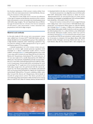
           preparations with knife -edge margins.             Val: 01/2024) (Figure 2) using a milling unit (Zirkonzahn M5
                                                              Heavy Metal, Zirkonzahn GmbH, Austria), as well as a replica of
                                                              the dental preparation in a cobalt-chrome alloy (Zirkonzahn
           Material and methods                               Sintermetall, Zirkonzahn GmbH, Austria), which was used for
                                                              mechanical testing (Figure 3). The internal surface of each crown
           In this pilot study, 20 full -contour resin nanoceramic crowns   was sandblasted with 50 -µm aluminum oxide powder (1.5 bar)
           were milled from Cerasmart270 CAD/CAM blocks and ran-  for 10 seconds at a distance of 10 mm (Basic classic 2945 -2026,
                                    ™
           domly divided into two experimental groups. This material is   Renfert GmbH, Hilzingen, Germany). Before each test, the co-
           a high -density composite resin containing a polymeric ma-  balt-chrome alloy die was cleaned with a wet compress and
           trix with the addition of silica nanoparticles of up to 1 µm (18)
           and barium glass (77% by weight).
             A dental preparation for a full -contour crown was per-
           formed on a lower right second premolar typodont tooth
           mounted on a dental anatomical model (ANA -4, Frasaco
                ®
           GmbH , Germany) (Figure 1). Axial guiding grooves were made
           1 -mm deep with a round end taper diamond bur (FG G850.010,
           Edenta AG. Hauptstrasse, Switzerland). The axial reduction of
           1.5 mm was made with a needle diamond bur (FG G859L.016,
           Edenta AG. Hauptstrasse, Switzerland) and the occlusal reduc-
           tion of 2.0 mm with a football diamond bur (FG G368.023, Eden-
           ta AG, Hauptstrasse, Switzerland) while maintaining the tooth
           morphology. The 45º -angle bevel of the functional cusp and the
           knife -edge margin were also performed with the needle dia-
           mond bur. The dental preparation was finished and polished
           with an Arkansas stone (Abrasive 025, Edenta AG. Hauptstrasse,
           Switzerland) followed by a fine -grain polishing rubber point
           (Exa Intrapol 050, Edenta AG. Hauptstrasse, Switzerland) to
           smooth the preparation’s surface. The whole procedure was   Figure 2. Full -contour crown milled from Cerasmart270™
           executed using rotary instruments with constant water cooling   resin nanoceramic CAD/CAM blocks (GC Corporation,
                                                               Tokyo, Japan).
           (Bora LK 1600732 -001, Bien -Air Dental, Bienne, Switzerland; CA


            Figure 1. Dental preparation for a full -contour crown with   Figure 3. Cobalt -chrome alloy die (Zirkonzahn
            a knife -edge margin, buccal view.                 Sintermetall, Zirkonzahn GMBH, Austria).

