


Page 28 - SPEMD_62-3
P. 28
154                    rev port estomatol med dent cir maxilofac. 2021;62(3):150-156


                                                              mented with zinc phosphate.The mean values of fracture re-
                                                              sistance reported were higher than those in the present study.
                                                              The differences in composition between the composite resin
                                                                                              ™
                                                              studied by Attia et al.  and Cerasmart270  affect the modulus
                                                                              22
                                                              of elasticity and flexural strength of these materials, which are

                                                              properties that influence their fracture resistance. It should
                                                              also be noted that Attia et al. used dentin as a substrate as
                                                              opposed to a cobalt -chrome alloy.
                                                                              16
                                                                 Wheyrauch et al.  analyzed the influence of the type of
                                                              cement on the fracture resistance of full -contour crowns
                                                              milled from CAD/CAM blocks of various ceramics and a nano-
                                                              -hybrid indirect composite resin. Several types of cement were
                                                              used, and they found no correlation between the two variables.
                                                              Differences between the materials’ compositions may be re-
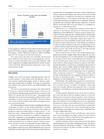
            Figure 5. Bar chart for fracture resistance mean values   sponsible for the incompatibility of their results with those of
                                                                                        23
            and standard deviation, in Newtons.               the present study. Nakamura et al.  also studied the influence
                                                              of various types of cement on the fracture resistance of full-
                                                              -contour zirconia crowns and found no significant differences

           tically significant differences were observed between the two   between groups.This result may be due to the high strength
           groups (p<0.001), as group 1 registered a mean value higher   and hardness inherent to that material. 24
           than group 2, with a difference of 866.4 N, which translates   There are limitations to this in vitro study. Instead of pre-
           into a 207.3% increase in the fracture resistance.  pared natural teeth, a single cobalt -chrome alloy die was used
             All crowns failed and were not repairable. All fractures   for the cementation of all 20 crowns tested. The sandblasting
           were confined to the crowns, and none progressed to the die.   can be speculated to have caused dimensional alterations in
           Group 1 crowns fractured at the crown -die interface, with   the die, increasing the cement gap for the next cementations
           crown fragments completely separating from the die. Group 2   and, therefore, influencing the fracture resistance and reduc-
           crowns failed by chipping without separation of the crown   ing standardization. Considering a potential actual increase of
           material from the die.                             cement gap, recent studies using CAD/CAM milled crowns
                                                              from  both  conventional  ceramics  and  resin  nanoceramics
                                                              showed that different cement thicknesses had no significant
           Discussion                                         effect on fracture resistance. 25,26  Furthermore, studies with
                                                              similar methodology also had standard deviations similar to
           A higher mean value and greater standard deviation were ob-  the ones of the present study. 15,16,20,23  Thus, there is no clear
           tained in group 1. Cementation with self -adhesive resin ce-  evidence that the standardization was reduced.
           ment showed significantly higher  results of fracture resist-  Nevertheless, the sandblasting caused micro -mechanical
           ance than cementation with RMGI. Thus, the null hypothesis   retentions on the die’s surface. In addition, the moduli of elas-
           was rejected. The mode of failure also differed between the   ticity of cobalt -chrome and dentin are different, which may
           two groups.                                        have led to results different than expected. Thus, it would be
             The mean values of fracture resistance were 1284.3±340.19   pertinent to use dentin as a substrate and one die per cement-
           N for group 1 and 417.9±106.35 N for group 2. Ferrario et al.   ed crown.
           noted that the average maximum bite force in premolars was   Another possible limitation is the use of putty to stabilize
                                           21
           206.01 N in women and 291.36 N in men.  The present study   the crown and die set. Being a flexible material, it may have
           obtained a minimum fracture resistance value of 285N, corre-  allowed some movement even though the crown and die set
           sponding to a crown cemented with RMGI. Therefore, almost   was firmly fixed to the test machine. However, the paper chart
           every crown withstood higher loads than the average bite forc-  from the software of the testing machine, in all specimens,
           es in the premolar area.                           demonstrated a linear curve until fracture occurred. Therefore,
             Notwithstanding the lack of studies regarding the influence   there is no indication that the putty elastomer impacted the
           of the type of cement on the fracture resistance of crowns   applied force or influenced the results obtained, but less flex-
           made from the resin nanoceramic featured in this study, it is   ible materials, such as gypsum or epoxy resin, could have been
           possible to verify concordant results in similar papers. 14,15  Lim   a more suitable alternative.
           and Lee’s study  demonstrated that a self -adhesive cement   The crowns cemented with self -adhesive resin cement
                       14
           increased the fracture resistance of an indirect composite res-  were tested immediately after light -curing. Thus, there was no
           in by 29%, compared to zinc phosphate. However, they regis-  post -curing time, which is described as increasing resin ce-

           tered mean values significantly lower than those of the present   ment’s properties, namely surface hardness. 27-29
           study, presumably due to using rectangular blocks as speci-  Another limitation of this study is the lack of cyclic and/or
           mens instead of crowns with adequate marginal adaptation.  thermomechanical load since restorations in the clinical en-
                     15
             Attia et al.  demonstrated that full -contour crowns milled   vironment fracture mainly due to fatigue. 30-33  Given the afore-
           from indirect composite resin CAD/CAM blocks and cemented   mentioned limitations, the direct transfer of results to in vivo
           with RMGI showed superior fracture resistance than those ce-  conditions is limited.

