
Page 34 - SPEMD_60-4
P. 34
182 rev port estomatol med dent cir maxilofac. 2019;60(4):175-188
This class is usually defined as a severely atrophic maxilla skillful in this technique and should be considered as the last
and is the most challenging class in full-arch rehabilitation. option in treatment planning due to the possible surgical
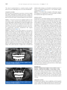
complications (Figure 11).
therapeutic options Option C – The severe bone resorption of this class requires
Both the anterior and posterior areas have a minimum height short implants or augmentation of the premaxilla to stabilize
and width, challenging the implant placement. Despite the the implants and support an overdenture.
different graft extension, the two fixed full-arch rehabilitation
options proposed require horizontal regeneration procedures. deciding factor
The key factor in deciding between these two approaches is
Option A – Placement of six or more straight implants at the whether the density of the remaining bone provides enough
same time or after a bilateral sinus lift procedure and hori- implant primary stability because both the previously men-
zontal regeneration. These implants are placed straight in the tioned options A and B are predictable. Therefore, two surgi-
region of the sinus lift graft, usually with entry points corre- cal protocols are suggested for a fixed full-arch rehabilitation
sponding to the canine, first premolar and first molar posi- depending on either a grafting or a graftless approach. How-
tions. Due to extensive horizontal bone resorption, a horizon- ever, in terms of the height level of atrophy, both techniques
tal augmentation procedure complements the surgical are sensitive to the patient’s preference, the risks involved,
rehabilitation scheme (Figure 10). the expertise and the learning curve of the surgeon. Patient
Option B – Placement of four short implants in the anterior risk factors such as heavy smoking, diabetes, bruxism, sinus
region of the maxilla. Two straight implants are placed in the pathology and other compromising factors have to be con-
lateral incisors position and two implants adjacent to the sidered in the decision process. Regardless of the vertical
maxillary sinus lift wall. In the posterior region of the maxilla, augmentation with sinus lift, treatment complexity increas-
two zygomatic implants are placed tilted forward to obtain es in the presence of a thin ridge (<5 mm). An alveolar width
implant anchorage and stability in the zygomatic bone by in- deficiency of this magnitude is often associated with loss of
creasing the implant length to ≥30 mm. Immediate loading is buccal cortical and/or medullary bone, compromising the pa-
possible if the anterior implants are stable. When it is not tients’ facial profiles and adding complexity to the treatment
possible to place stable implants in the anterior region, four plan. In these cases, a horizontal ridge augmentation to
39
zygomatic implants can be used. The main advantage of this cover the buccal surface of every implant and re-establish
option is allowing immediate loading without a grafting pro- the patients’ anatomy is mandatory. Option B with zygomat-
cedure. This option requires a surgeon who is trained and ic implants is one of several techniques described in the liter-
ature to approach the atrophic maxilla, with several prospec-
tive studies reporting successful outcomes as well as
recognized advantages such as reduced treatment time, de-
creased morbidity and a smaller number of implants neces-
sary to support fixed prostheses. 40,41 Several studies have
evaluated the use of zygomatic implants combined with
standard implants for immediate loading and have reported
a high survival rate of 95.8% to 100%, which implies that zy-
gomatic implants may be used with immediate function pro-
tocols. 42-44 Despite the high survival rate reported in the liter-
ature, attention should be drawn to the decision-making
process due to the risk of intra- or post-operative complica-
tions involved: infections/sinusitis in the maxillary sinus, in-
Figure 10. Example of Maxilla CC Class V, Option A and traoral soft-tissue problems, oroantral fistula, orbital injury
Mandible CC Class IV, Option A
and intracranial penetration. 40
Mandible CCI
Anterior – Available bone (height >16 mm; width >6 mm)
Posterior – Available bone (height >12 mm; width >6 mm)
In the anterior region of the mandible, the bone height meas-
ured from the osteotomy level, with a >6-mm crestal width,
to the inferior border of the mandible is >16 mm. In the pos-
terior region, the distance from the >6-mm alveolar crestal
width to a 2-mm safe distance from the mandibular canal is
>12 mm.
therapeutic options
Considering the posterior bone availability and the favorable
Figure 11. Example of Maxilla CC Class V, Option B and anterior region with a higher density bone, two fixed full-arch
Mandible CC Class IV, Option A
options and two removable full-arch schemes are proposed.