


Page 52 - SPEMD_60-3
P. 52
140                    rev port estomatol med dent cir maxilofac. 2019;60(3):137-144


            Figure 5. Post -operative radiograph               Figure 6. 19 -month follow -up


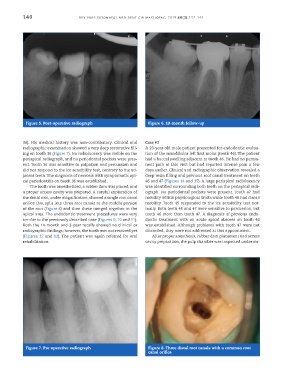
           36). His medical history was non -contributory. Clinical and   Case #3
           radiographic examination showed a very deep restorative fill-  A 28 -year -old male patient presented for endodontic evalua-
           ing on tooth 36 (Figure 7). No radiolucency was visible on the   tion of the mandibular left first molar (tooth 46). The patient
           periapical radiograph, and no periodontal pockets were pres-  had a buccal swelling adjacent to tooth 46. He had no perma-
           ent. Tooth 36 was sensitive to palpation and percussion and   nent pain at this visit but had reported intense pain a few
           did not respond to the ice sensibility test, contrary to the ad-  days earlier. Clinical and radiographic observation revealed a
           jacent teeth. The diagnosis of necrosis with symptomatic api-  deep resin filling and previous root canal treatment on teeth
           cal periodontitis on tooth 36 was established.     46 and 47 (Figures 14 and 15). A large periapical radiolucency
             The tooth was anesthetized, a rubber dam was placed, and   was identified surrounding both teeth on the periapical radi-
           a proper access cavity was prepared. A careful exploration of   ograph. No periodontal pockets were present. Tooth 47 had
           the distal root, under magnification, showed a single root canal   mobility within physiological limits while tooth 46 had class I
           orifice that split into three root canals at the middle portion   mobility. Tooth 45 responded to the ice sensibility test nor-
           of the root (Figure 8) and then these merged together at the   mally. Both teeth 46 and 47 were sensitive to percussion, but
           apical area. The endodontic treatment procedures were very   tooth 46 more than tooth 47. A diagnosis of previous endo-
           similar to the previously described case (Figures 9, 10 and 11).   dontic treatment with an acute apical abscess on tooth 46
           Both the 14 -month and 3 -year recalls showed no clinical or   was established. Although problems with tooth 47 were not
           radiographic findings; however, the tooth was not restored yet   discarded, they were not addressed at this appointment.
           (Figures 12 and 13). The patient was again referred for oral   After proper anesthesia, rubber dam placement and access
           rehabilitation.                                    cavity preparation, the pulp chamber was inspected under mi-


            Figure 7. Pre -operative radiograph                Figure 8. Three distal root canals with a common root
                                                               canal orifice

