


Page 54 - SPEMD_60-3
P. 54
142                    rev port estomatol med dent cir maxilofac. 2019;60(3):137-144


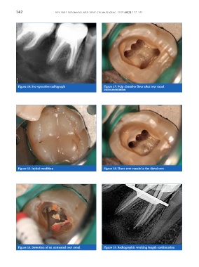
            Figure 14. Pre -operative radiograph               Figure 17. Pulp chamber floor after root canal
                                                               instrumentation


            Figure 15. Initial condition                       Figure 18. Three root canals in the distal root


            Figure 16. Detection of an untreated root canal    Figure 19. Radiographic working length confirmation

