


Page 53 - SPEMD_60-3
P. 53
rev port estomatol med dent cir maxilofac . 2019;60(3):137-144         141


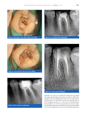
            Figure 9. Obturation of the three distal root canals  Figure 12. 14 -month recall radiograph


            Figure 10. Pulp chamber after root canal filling


                                                                Figure 13. 3 -year recall radiograph


                                                               croscope. Four main root canals (two mesial and two distal)
                                                               with gutta -percha filling were found, as well as a fifth inde-
                                                               pendent root canal (distobuccal) that had not been instru-
                                                               mented (Figure 16). The distal root had a Gulabivala Type 12
                                                               (3 -2) configuration (Figures 17 and 18). All root canals were
                                                               instrumented to an F2 ProTaper rotary file. A rigorous irriga-
            Figure 11. Post -operative radiograph              tion protocol was performed, and the root canals were dressed
                                                               with calcium hydroxide. At the second visit, three weeks later,

