


Page 51 - SPEMD_60-3
P. 51
rev port estomatol med dent cir maxilofac . 2019;60(3):137-144         139


           hol. The canals orifices were sealed with flowable composite   chamber floor with a DG16 Endo Explorer, three main root ca-
           resin. The access cavities were temporarily restored, and the   nals (two mesial and one distal) with gutta -percha filling were
           patients were referred for coronal rehabilitation.  found, and a careful analysis of the area surrounding the dis-
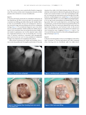
                                                               tal canal revealed two untreated canals in the distal root (dis-
           Case #1                                             tobuccal and distolingual root canals) (Figure 2). The three root
           A 34 -year -old female presented for endodontic evaluation of   canals had three distinct root canal orifices but merged togeth-
           her mandibular left first molar (tooth 46). The patient’s chief   er in the apical area, presenting a Gulabivala Type 9 (3 -1) con-
           complaint was feeling pain when chewing. Her medical histo-  figuration. After root canal negotiation and working length
           ry was noncontributory. Clinical and radiographic examina-  measurement, rotary instrumentation was performed to an F2
           tion revealed a large and deep filling and previous endodontic   ProTaper file. The root canals were disinfected, and a calcium
           therapy (Figure 1). The periodontal ligament space was wider   hydroxide dressing was used between visits. In the second
           than in normal conditions. Tooth mobility was within physio-  visit, a new disinfection protocol was performed, and the root
           logical limits, and there were no periodontal pockets. Tooth 46   canal  obturation  was  completed (Figures  3,  4  and  5). The
           was tender to percussion, but not the adjacent teeth, which   19 -month follow -up showed no clinical or radiographic patho-
           also responded normally to the ice sensibility test. The diag-  logical findings (Figure 6).
           nosis  of  previous  endodontic  treatment  with  symptomatic
           apical periodontitis on tooth 46 was established. Endodontic   Case #2
           therapy was proposed and accepted by the patient.   A 36 -year -old male patient came to an emergency visit with a
              After proper anesthesia and rubber dam isolation, the ac-  chief complaint of feeling permanent pain that increased
           cess cavity was performed. During the exploration of the pulp   when chewing with the mandibular right first molar (tooth


            Figure 1. Pre -operative radiograph                 Figure 3. Working length measurement


            Figure 2. Pulp chamber floor showing three root canals   Figure 4. Final root canal filling
            in the distal root

