
Page 54 - SPEMD_59-1
P. 54
46 rev port estomatol med dent cir maxilofac. 2018;59(1):44-48
screws of the 2.0-mm system by intraoral access; the bilateral
subcondylar fractures were conservatively treated.
The patient was kept without IMF for 24 hours. After this
period wearing the orthodontic appliance and tooth syntheses,
two rubber appliances with nearly 5-mm height were placed on
the posterior occlusal region bilaterally, and IMF was performed
with elastics involving primarily the anterior teeth. This fixation
associated with posterior support was kept for two days.
After this period, the patient returned to the dentist’s offi-
ce and the posterior support was removed. The intermaxillary
fixation with elastics was kept for seven days further.
After removal of the intermaxillary fixation, the patient
was referred to postoperative physiotherapy, including ope-
ning, bilateral laterality and protrusion exercises, of which the
last two were performed with the help of manual force. The
number of repetitions of the exercises was 40 times for ope-
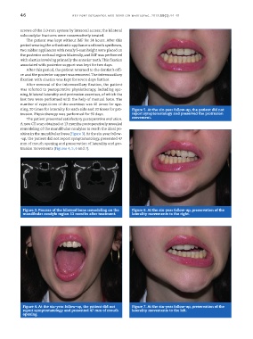
ning, 20 times for laterality for each side and 20 times for pro- Figure 5. At the six-year follow-up, the patient did not
trusion. Physiotherapy was performed for 90 days. report symptomatology and preserved the protrusion
The patient presented satisfactory postoperative evolution. movement.
A new CT scan obtained at 13 months postoperatively revealed
remodeling of the mandibular condyles to reach the ideal po-
sition in the mandibular fossa (Figure 3). At the six-year follow-
-up, the patient did not report symptomatology, presented 47
mm of mouth opening and preservation of laterality and pro-
trusion movements (Figures 4, 5, 6 and 7).
Figure 3. Process of the bilateral bone remodeling on the Figure 6. At the six-year follow-up, preservation of the
mandibular condyle region 13 months after treatment. laterality movements to the right.
Figure 4. At the six-year follow-up, the patient did not Figure 7. At the six-year follow-up, preservation of the
report symptomatology and presented 47 mm of mouth laterality movements to the left.
opening.