


Page 30 - SPEMD_67-1
P. 30
24                      rev port estomatol med dent cir maxilofac . 2026;67(1):21-26


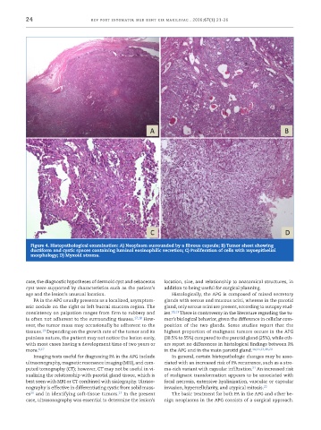
            Figure 4. Histopathological examination: A) Neoplasm surrounded by a fibrous capsule; B) Tumor sheet showing
            ductiform and cystic spaces containing luminal eosinophilic secretion; C) Proliferation of cells with myoepithelial
            morphology; D) Myxoid stroma.


           case, the diagnostic hypotheses of dermoid cyst and sebaceous   location, size, and relationship to anatomical structures, in
           cyst were supported by characteristics such as the patient’s   addition to being useful for surgical planning.
           age and the lesion’s unusual location.                Histologically, the APG is composed of mixed secretory
             PA in the APG usually presents as a localized, asymptom-  glands with serous and mucous acini, whereas in the parotid
           atic nodule on the right or left buccal mucosa region. The   gland, only serous acini are present, according to autopsy stud-
           consistency on palpation ranges from firm to rubbery and   ies. 18,21  There is controversy in the literature regarding the tu-
           is often not adherent to the surrounding tissues. 17,18  How-  mor’s biological behavior, given the difference in cellular com-
           ever, the tumor mass may occasionally be adherent to the   position of the two glands. Some studies report that the
                 19
           tissues.  Depending on the growth rate of the tumor and its   highest proportion of malignant tumors occurs in the APG
           painless nature, the patient may not notice the lesion early,   (38.5% to 55%) compared to the parotid gland (25%), while oth-
           with most cases having a development time of two years or   ers report no differences in histological findings between PA
           more. 6,17                                         in the APG and in the main parotid gland. 14,15,17,19,20
             Imaging tests useful for diagnosing PA in the APG include   In general, certain histopathologic changes may be asso-
           ultrasonography, magnetic resonance imaging (MRI), and com-  ciated with an increased risk of PA recurrence, such as a stro-
                                                                                               21
           puted tomography (CT); however, CT may not be useful in vi-  ma-rich variant with capsular infiltration.  An increased risk
           sualizing the relationship with parotid gland tissue, which is   of malignant transformation appears to be associated with
           best seen with MRI or CT combined with sialography. Ultraso-  focal necrosis, extensive hyalinization, vascular or capsular
           nography is effective in differentiating cystic from solid mass-  invasion, hypercellularity, and atypical mitosis. 22
                                             17
           es  and in identifying soft-tissue tumors.  In the present   The basic treatment for both PA in the APG and other be-
            20
           case, ultrasonography was essential to determine the lesion’s   nign neoplasms in the APG consists of a surgical approach.

