


Page 29 - SPEMD_67-1
P. 29
rev port estomatol med dent cir maxilofac . 2026;67(1):21-26             23


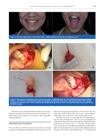
             Figure 2. Extraoral appearance of the lesion with a slight swelling just below the zygomatic arch.


             Figure 3. Intraoperative sequence of the transoral excision: A) Identification of a well-defined, encapsulated nodular
             lesion in the accessory parotid gland region; B) Complete enucleation of the lesion with preservation of the capsule
             integrity; C) Excision of the entire accessory parotid gland along with the lesion; D) Repositioning of the buccal fat pad
             (Bichat’s fat pad).


              Informed consent and authorization for the use of the pre-  first step in diagnosing pathologies in this structure and, con-
           sented clinical data were obtained to ensure the patient’s con-  sequently, in choosing the best way to treat tumors satisfac-
           fidentiality and anonymity, in accordance with applicable   torily and safely, to avoid recurrences and complications.
           ethical guidelines.                                   The APG can serve as a potential site for benign and ma-
                                                               lignant lesion development, with PA and mucoepidermoid
                                                               carcinoma as the most common benign and malignant lesions,
           Discussion and conclusions                          respectively, in this structure. 6,14-16  However, various lesions
                                                               may be considered in the differential diagnosis of APG pathol-
           PA in the APG is a relatively uncommon condition, scarcely   ogies, including dermoid cyst, schwannoma, lipoma, primary
           reported in the literature. Awareness of APG’s existence is the   Stensen’s duct tumor, and neurofibroma. 17,18  In the present

