


Page 34 - SPEMD_67-1
P. 34
28                      rev port estomatol med dent cir maxilofac . 2026;67(1):27-33


           oral lesions present as asymptomatic, well-demarcated exo-
           phytic growths with superficial papillary or verruciform pro-
           jections, often resembling cauliflower. The lesions are usually
           white but can appear pink or erythematous. 9
             VC accounts for approximately 2−12% of all oral carcino-
              10
           mas.  Although the etiology is not well understood, risk fac-
           tors commonly associated with  VC development include
           smoking, tobacco chewing, alcohol consumption, and poor
           oral hygiene. Other etiological factors include human papillo-
           mavirus (HPV), areca nut chewing, immunosuppression, oral
           leukoplakia, and oral submucous fibrosis. 11,12  The treatment
                                                         13
           of choice for oral VC is surgical excision with wide margins.
           Overall, the prognosis is favorable, with a 5-year survival rate
           of approximately 80%; however, the recurrence rate varies
           from 30% to 50%. 14,15
             This paper presents two cases of oral VC in elderly men
           occurring in unusual locations — the palate and the lower lip,   Figure 2. Photomicrograph showing exophytic tumor
           followed by a literature review emphasizing clinical features,   with church spire sign and broad, bulbous epithelial
           histological findings, and treatment modalities.     ridges that appeared to push against the underlying
                                                                connective tissue (H&E, 40x).

           Case reports

           Case 1
           A 77-year-old male patient was referred to the (placeholder for
           the institution’s name) for evaluation of a white verrucous le-
           sion on the lower lip with a duration of approximately 1 year.
           The medical history revealed no systemic diseases or drug
           allergies. In addition, the patient reported a 63-year history of
           smoking. On physical examination, no regional lymphadenop-
           athy was observed. A whitish exophytic lesion was present on
           the vermilion border and mucosa of the lower lip. The plaque
           was thick and well-circumscribed, with a papillary surface and
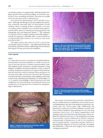
           a sessile base, measuring approximately 3 cm in diameter (Fig-
           ure 1). Based on these findings, a clinical hypothesis of VC was
           established. An incisional biopsy was performed under local
           anesthesia, and the specimen was submitted for histopatho-
           logical examination.
                                                                Figure 3. Photomicrograph showing mild cytologic
                                                                atypia in the basal layer and absence of frank invasion
                                                                (H&E, 100x).


                                                                 Microscopic examination demonstrated a well-differen-
                                                              tiated stratified squamous epithelium with marked superfi-
                                                              cial keratinization and verrucous exophytic projections, con-
                                                              sistent  with  the  “church  spire”  sign.  Additionally, the
                                                              hyperplastic epithelium exhibited endophytic growth with
                                                              broad, bulbous epithelial ridges pushing into the underlying
                                                              connective tissue (Figure 2). At higher magnification, mild
                                                              cytologic atypia was observed in the basal layer, with no ev-
                                                              idence of overt invasion (Figure 3). The underlying connective
                                                              tissue showed a chronic inflammatory infiltrate composed
                                                              predominantly of lymphocytes, as well as rich vascularity
                                                              and no lymphovascular or perineural invasion. Therefore, a
                                                              histopathologic diagnosis of VC was confirmed. The patient
            Figure 1. Extraoral examination showing a white
            verrucous lesion on the lower lip.                was referred to a head and neck surgeon and is currently
                                                              undergoing treatment.

