


Page 28 - SPEMD_67-1
P. 28
22                      rev port estomatol med dent cir maxilofac . 2026;67(1):21-26


           scribed as “soft-tissue masses” located in the middle of the buc-  oral clinical examination revealed the presence of a palpable
           cal mucosa, usually unilateral, without painful symptoms, slow   nodule, both intra- and extraoral, located in the buccal mu-
           growing, and sessile due to the fixation to the masseteric fascia. 6  cosa, which was firm, mobile, asymptomatic, and well-cir-
             Pleomorphic adenoma (PA) is considered the most com-  cumscribed. No lymph node enlargement was found in the
           mon salivary gland neoplasm. It manifests in all age groups,   lateral cervical chains, nor in the submandibular or parotid
           with the most frequent involvement in the 30 to 60-year age   chains.
           group and a slight predilection for women. 7,8  PA occurs most   Based on the diagnostic hypotheses of a dermoid cyst and
           commonly in the parotid gland — 40 to 75%, followed by the   a sebaceous cyst, enucleation of the mass was performed. The
           submandibular gland and minor salivary glands. 9,10  The palate   surgical procedure was executed under local infiltrative anes-
           is the most common intraoral site, mainly the hard palate,   thesia with 2% articaine. The intraoperative aspiration biopsy
           while occurrences in other intraoral sites include the upper lip   was negative, indicating a solid lesion. An intraoral incision
           and buccal mucosa. 11                              was initiated superiorly to the papilla of the parotid duct. Care-
             As in the main parotid gland, PA is the most common be-  fully, continuous blunt dissection (divulsion) was performed
           nign tumor of the APG. Clinically, it usually presents as a firm,   meticulously to preserve the surrounding soft tissues (Figure
           slowly progressive, and painless unilateral swelling, typically   3A). Intraoperatively, a nodular and encapsulated lesion was
           without facial nerve involvement. 5,12,13  However, data on the   observed. This mass was characterized by well-defined borders
           prevalence of PA in the APG are scarce in the literature. There-  and was readily separable from the adjacent tissues. The in-
           fore, the present study aims to elucidate a clinical case of PA   traoperative location was highly suggestive of a development
           in the APG in a young patient.                     arising from the APG. The lesion was entirely removed, ensur-
                                                              ing the integrity of the lining capsule was strictly preserved
                                                              (Figure 3B). Along with the tumor, the entire APG was also ex-
           Case report                                        cised (Figure 3C). After the complete excision, the buccal fat
                                                              pad (Bichat’s fat pad) was repositioned to mitigate potential
           A 20-year-old female, systemically healthy, was referred to the   facial asymmetry (Figure 3D). Primary closure was subsequent-
           stomatology service complaining of a swelling in the right   ly achieved using a single stitch suture.
           cheek. It had been progressively increasing for approximately   The histopathological analysis showed a benign salivary
           18 months, without painful symptoms, but with aesthetic dis-  gland neoplasm with an encapsulated appearance, character-
           comfort. Ultrasonography revealed a rounded hypoechoic im-  ized by the proliferation of epithelial cells forming ductal and
           age, without associated vascularization, compatible with a   cystic spaces, and myoepithelial cells of varying morphology,
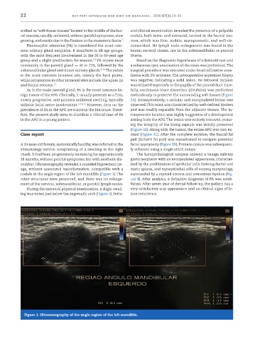
           nodule in the angle region of the left mandible (Figure 1). The   surrounded by a myxoid stroma and sometimes hyaline (Fig-
           other structures were preserved, and there was no enlarge-  ure 4). After analysis, a definitive diagnosis of PA was estab-
           ment of the cervical, submandibular, or parotid lymph nodes.  lished. After seven year of dental follow-up, the patient has a
             During the extraoral physical examination, a slight swell-  very satisfactory scar appearance and no clinical signs of le-
           ing was noted just below the zygomatic arch (Figure 2). Intra-  sion recurrence.


            Figure 1. Ultrasonography of the angle region of the left mandible.

