
Page 60 - SPEMD_61-3
P. 60
150 rev port estomatol med dent cir maxilofac. 2020;61(3):148-153
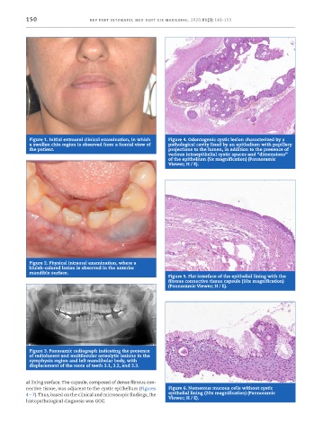
Figure 1. Initial extraoral clinical examination, in which Figure 4. Odontogenic cystic lesion characterized by a
a swollen chin region is observed from a frontal view of pathological cavity lined by an epithelium with papillary
the patient. projections to the lumen, in addition to the presence of
various intraepithelial cystic spaces and “dimensions”
of the epithelium (5x magnification) (Pannoramic
Viewer; H / E).
Figure 2. Physical intraoral examination, where a
bluish -colored lesion is observed in the anterior
mandible surface.
Figure 5. Flat interface of the epithelial lining with the
fibrous connective tissue capsule (10x magnification)
(Pannoramic Viewer; H / E).
Figure 3. Panoramic radiograph indicating the presence
of radiolucent and multilocular osteolytic lesions in the
symphysis region and left mandibular body, with
displacement of the roots of teeth 3.1, 3.2, and 3.3.
al lining surface. The capsule, composed of dense fibrous con-
nective tissue, was adjacent to the cystic epithelium (Figures Figure 6. Numerous mucous cells without cystic
4 – 7). Thus, based on the clinical and microscopic findings, the epithelial lining (20x magnification) (Pannoramic
Viewer; H / E).
histopathological diagnosis was GOC.