
Page 54 - SPEMD_60-4
P. 54
202 rev port estomatol med dent cir maxilofac. 2019;60(4):197-204
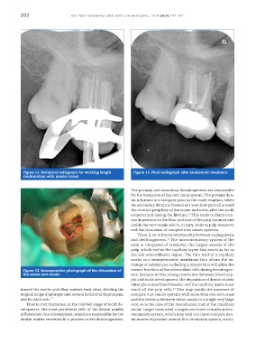
Figure 12. Periapical radiograph for working length Figure 14. Final radiograph after endodontic treatment
confirmation with master cones
The primary and secondary dentinogenesis are responsible
for the formation of the root canal system. The primary den-
tin is formed at a fast pace prior to the tooth eruption, while
the secondary dentin is formed at a very slow pace all around
the internal periphery of the crown and roots, after the tooth
13
eruption and during the lifetime. This leads to dentin ma-
trix deposition on the floor and roof of the pulp chamber and
inside the root canals which, in turn, leads to pulp recession
and the formation of complex root canals systems.
There is an intimate relationship between angiogenesis
and dentinogenesis. The microcirculatory system of the
13
pulp is composed of arterioles, the largest vessels of the
pulp, which end in the capillary layers that reach as far as
the sub-odontoblastic region. The thin wall of a capillary
works as a semipermeable membrane that allows the ex-
change of substances, including nutrients that will allow the
Figure 13. Intraoperative photograph of the obturation of correct function of the odontoblast cells during dentinogen-
the seven root canals esis. Because of this strong correlation between blood sup-
ply and tooth development, the deposition of dentin matrix
takes place near blood vessels, and the capillary layers must
toward the center until they contact each other, dividing the reach all the pulp cells. This may justify the presence of
14
original single diaphragm into several horizontal diaphragms, complex root canals systems with more than one root canal
one for each root. 12 and the isthmus between these canals in a single very large
Prior to root formation, at the late bell stage of tooth de- root, as is the case of the mesiobuccal root of the maxillary
velopment, the most peripheral cells of the dental papilla molar. Larger roots need a larger and more complex micro-
differentiate into odontoblasts, which are responsible for the circulatory system, which may lead to a more complex den-
dentin matrix secretion in a process called dentinogenesis. tin matrix deposition around this circulatory system, result-