
Page 52 - SPEMD_60-4
P. 52
200 rev port estomatol med dent cir maxilofac. 2019;60(4):197-204
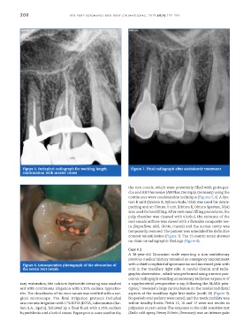
Figure 5. Periapical radiograph for working length Figure 7. Final radiograph after endodontic treatment
confirmation with master cones
the root canals, which were posteriorly filled with gutta-per-
cha and AH Plus sealer (AH Plus, Dentsply, Germany) using the
continuous wave condensation technique (Figures 5, 6). A Sys-
tem B unit (System B, Sybron Endo, USA) was used for down-
packing and an Obtura II unit (Obtura II, Obtura Spartan, USA)
was used for backfilling. After root canal filling procedures, the
pulp chamber was cleaned with alcohol, the entrance of the
root canals orifices was closed with a flowable composite res-
in (Supraflow, R&S, CFPM, France) and the access cavity was
temporarily restored. The patient was scheduled for definitive
coronal rehabilitation (Figure 7). The 15-month recall showed
no clinic or radiographic findings (Figure 8).
Case # 2
A 38-year-old Caucasian male reporting a non-contributory
previous medical history attended an emergency appointment
Figure 6. Intraoperative photograph of the obturation of with a chief complaint of spontaneous and increased pain with
the seven root canals cold in the maxillary right side. A careful clinical and radio-
graphic observation , which was performed using a recent pan-
oramic radiograph avoiding unnecessary radiation exposure of
rary restoration, the calcium hydroxide dressing was washed a supplemental preoperative x-ray, following the ALARA prin-
11
out with continuous irrigation with 5.25% sodium hypochlo- ciples, revealed a large carious lesion on the mesial and distal
rite. The cleanliness of the root canals was verified with a sur- aspects of the maxillary right first molar (tooth 16) (Figure 9).
gical microscope. The final irrigation protocol included No periodontal pockets were noted, and the tooth mobility was
one-minute irrigation with 17% EDTA (EDTA, Laboratorios Clar- within healthy limits. Teeth 15, 16 and 17 were not tender to
ben S.A., Spain), followed by a final flush with 5.25% sodium palpation or percussion. The response to the cold-sensitive test
hypochlorite and alcohol rinses. Paper points were used to dry (Endo cold spray, Henry Schein, Germany) was an intense pain