
Page 53 - SPEMD_60-4
P. 53
rev port estomatol med dent cir maxilofac . 2019;60(4):197-204 201
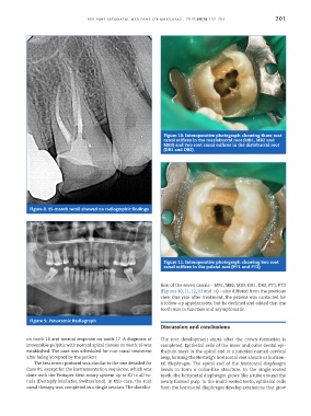
Figure 10. Intraoperative photograph showing three root
canal orifices in the mesiobuccal root (MB1, MB2 and
MB3) and two root canal orifices in the distobuccal root
(DB1 and DB2)
Figure 8. 15-month recall showed no radiographic findings
Figure 11. Intraoperative photograph showing two root
canal orifices in the palatal root (PT1 and PT2)
tion of the seven canals – MB1, MB2, MB3, DB1, DB2, PT1, PT2
(Figures 10, 11, 12, 13 and 14) – also differed from the previous
case. One year after treatment, the patient was contacted for
a follow-up appointment, but he declined and added that the
tooth was in function and asymptomatic.
Figure 9. Panoramic Radiograph
Discussion and conclusions
on tooth 16 and normal response on tooth 17. A diagnosis of The root development starts after the crown formation is
irreversible pulpitis with normal apical tissues on tooth 16 was completed. Epithelial cells of the inner and outer dental epi-
established. The case was scheduled for root canal treatment thelium meet in the apical end at a junction named cervical
after being accepted by the patient. loop, forming the Hertwig’s horizontal root sheath or horizon-
The treatment protocol was similar to the one detailed for tal diaphragm. The apical end of the horizontal diaphragm
Case #1, except for the instrumentation sequence, which was bends to form a collar-like structure. In the single-rooted
done with the Protaper Next rotary system up to X2 in all ca- teeth, the horizontal diaphragm grows like a tube around the
nals (Dentsply Maillefer, Switzerland). In this case, the root newly formed pulp. In the multi-rooted teeth, epithelial cells
canal therapy was completed in a single session. The distribu- from the horizontal diaphragm develop extensions that grow