
Page 51 - SPEMD_60-4
P. 51
rev port estomatol med dent cir maxilofac . 2019;60(4):197-204 199
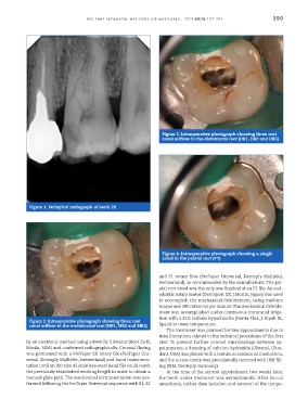
Figure 3. Intraoperative photograph showing three root
canal orifices in the distobuccal root (DB1, DB2 and DB3)
Figure 1. Periapical radiograph of tooth 26
Figure 4. Intraoperative photograph showing a single
canal in the palatal root (PT)
and F1 rotary files (ProTaper Universal, Dentsply Maillefer,
Switzerland), as recommended by the manufacturer. The pal-
atal root canal was the only one finished at an F2 file. An end-
odontic rotary motor (Dentaport ZX, J.Morita, Japan) was used
to accomplish the mechanical debridement, using medium
torque and 300 rotations per minute. The mechanical debride-
ment was accomplished under continuous intracanal irriga-
tion with 5.25% sodium hypochlorite (Denta Flux, J. Ripoll SL,
Figure 2. Intraoperative photograph showing three root
canal orifices in the mesiobuccal root (MB1, MB2 and MB3) Spain) at room temperature.
The treatment was planned for two appointments due to
time limitations related to the technical procedures of the first
by an electronic method using a Root Zx II locator (Root Zx II, visit. To prevent further coronal microleakage between ap-
Morita, USA) and confirmed radiographically. Coronal flaring pointments, a dressing of calcium hydroxide (Ultracal, Ultra-
was performed with a ProTaper SX rotary file (ProTaper Uni- dent, USA) was placed with a lentulo as intracanal medication,
versal, Dentsply Maillefer, Switzerland) and hand instrumen- and the access cavity was provisionally restored with IRM fill-
tation until an ISO size.15 stainless-steel hand file could reach ing (IRM, Dentsply, Germany).
the previously established working length in order to obtain a At the time of the second appointment, two weeks later,
manual glide path. The mechanical instrumentation was per- the tooth under treatment was asymptomatic. After buccal
formed following the ProTaper Universal sequence with S1, S2 anesthesia, rubber dam isolation and removal of the tempo-