
Page 17 - SPEMD_60-4
P. 17
rev port estomatol med dent cir maxilofac . 2019;60(4):163-168 165
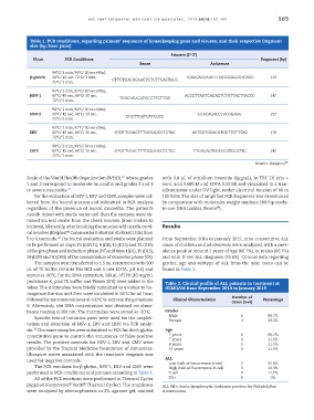
Table 1. PCR conditions, regarding primers’ sequence of housekeeping gene and viruses, and their respective fragment
size (bp, base pairs)
Primers (5’-3’)
Virus PCR Conditions Fragment (bp)
Sense Antisense
94°C/ 2 min, 94°C/ 30 sec (40x),
β-globin 60°C/ 45 sec, 72°C/ 1 min, TCACCACCAACTTCATCCAGCTTCACC 123
72°C/ 5 min. CTTCTGACACAACTGTGTTCACTAGC
94°C/ 2 min, 94°C/ 30 sec (40x),
HSV-1 60°C/ 45 sec, 68°C/ 30 sec, TGGGACACATGCCTTCTTGG ACCCTTAGTCAGACTCTGTTACTTACCC 147
72°C/ 5 min.
94°C/ 2 min, 94°C/ 30 sec (40x),
HSV-2 60°C/ 45 sec, 68°C/ 30 sec, CGCTTCATCATGGGC GTACAGACCTTCGGAGG 227
72°C/ 5 min.
94°C/ 2 min, 94°C/ 30 sec (40x),
EBV 60°C/ 45 sec, 68°C/ 30 sec, GTGTTCGACTTTGCCAGCCTCTAC ACTCGTGCACGTGCTTCTTTAC 176
72°C/ 5 min.
94°C/ 2 min, 94°C/ 30 sec (40x),
CMV 60°C/ 45 sec, 68°C/ 30 sec, GTGTTCGACTTTGCCAGCCTCTAC TTGACACTCGCGCATGCATTC 242
72°C/ 5 min.
19
Source: Adapted .
Scale of the World Health Organization (WHO), where grades with 0.8 μL of ethidium bromide (1μg/μL), in TBE 1X (tris +
16
1 and 2 correspond to moderate mucositis and grades 3 and 4 boric acid 0.089 M and EDTA 0.02 M) and visualized in a tran-
to severe mucositis. 17 silluminator under UV light, under electrical tension of 80 to
For the evaluation of HSV-1, EBV and CMV, samples were col- 100 Volts. The size of amplified PCR fragments was determined
lected from the buccal mucosa and submitted to PCR analysis by comparison with molecular weight markers (100 bp ready-
®
regardless of the presence of buccal mucositis. The patient’s to-use DNA Ladder, Bioron ).
mouth rinsed with sterile water and then the samples were ob-
tained via oral swabs from the cheek mucosa (from molars to
incisors), bilaterally, after brushing the mucosa with sterile cervi- Results
®
cal brushes (Kolplast Commercial Industrial do Brasil Ltda) from
10
5 to 10 seconds. The buccal evaluation and swabs were planned From September 2014 to January 2015, nine consecutive ALL
to be performed on days 0/1 (D0/D1), 8 (D8), 15 (D15) and 35 (D35) cases of children and adolescents were analyzed, with a prev-
of the pre-phase and induction phase (P/I) and days 1(D1), 15 (D15), alence peak at around 2 years of age (66.7%), in males (66.7%)
29 (D29) and 50 (D50) of the consolidation of remission phase (CR). and with B-cell ALL diagnosis (55.6%). Clinical data regarding
The samples were transferred to 1.5 mL microtubes with 500 gender, age and subtype of ALL from the nine cases can be
μL of TE buffer (10 mM Tris HCl and 1 mM EDTA, pH 8.0) and found in Table 2.
stored at -20ºC. For the DNA extraction, 500 μL of TPK (10 mg/mL
proteinase K, plus TE buffer and Tween 20%) were added to the Table 2. Clinical profile of ALL patients in treatment at
tubes. The microtubes were briefly submitted to a vortex to ho- HEMOAM from September 2014 to January 2015
mogenize the mix and then were incubated at 56°C for an hour,
followed by ten more minutes at 100°C to activate the proteinase Clinical Characteristics Number of Percentage
cases (n=9)
K. Afterwards, the DNA concentration was obtained via absor-
bance reading at 260 nm. The microtubes were stored at -20°C. Gender
Specific sets of initiation pairs were used for the amplifi- Male 6 3 66.7%
33.3%
Female
cation and detection of HSV-1, EBV and CMV via PCR analy-
19
sis. The same samples were submitted to PCR for the β-globin Age
constitutive gene to control the occurrence of false-positive 2 years 6 66.7%
results. The positive controls for HSV-1, EBV and CMV were 7 years 1 1 11.1%
9 years
11.1%
provided by the Tropical Medicine Foundation of Amazonas. 14 years 1 11.1%
Ultrapure water associated with the reaction’s reagents was
used for negative controls. ALL 5 55.6%
Low Risk of Recurrence B-cell
The PCR reactions for β-globin, HSV-1, EBV and CMV were High Risk of Recurrence B-cell 3 33.3%
performed in PCR conditions and primers according to Table 1. T-cell 1 11.1%
All of the PCR reactions were performed in Thermal Cycler PH+ 0 0%
(Applied Biosystems Veriti Thermal Cycler). The amplicons ALL PH+: Acute lymphocytic leukemia positive for Philadelphia
®
®
were analyzed by electrophoresis in 2% agarose gel, stained chromosome.