
Page 60 - SPEMD_60-3
P. 60
148 rev port estomatol med dent cir maxilofac. 2019;60(3):145-149
Unexpected morphological variations make endodontic
treatment a challenge for clinicians, who must be aware of all
possible anatomical configurations of the tooth as well as the
different diagnostic features that lead to successful endodon-
tic treatment. 12-14
Radiographic examinations are an essential component
in endodontic treatment. They are used for diagnosis, plan-
ning and to evaluate the success rate of treatments. The
amount of information obtained by two-dimensional periapi-
cal radiographs is limited, since it may be affected by geomet-
ric distortions and overlap of anatomical structures. 1,4,9,15 The
use of CT scans has been encouraged to diagnose cases of
multiple root canals. 3,16 In the present report, this resource
ensured treatment safety as it indicated the location of the
root canals. However, when computed tomography is not
available, radiographic images at different angulations can
be an option. 4,10
Using magnification might also be an advantage when
dealing with severe anatomic deviations. 6-8,17,18 In order to car-
ry out the root canal treatment of the presented case, an op-
erating microscope was used, at 16x and 25x magnification.
This device also contributed to the success of the technical
procedure, by preventing excessive wear of the dentin area and
improving visibility to locate the root canals.
In this case report, the possible etiology for pulp necrosis
and apical periodontitis in the teeth may have been an old
traumatic event reported by the patient.
The therapeutic protocol using tomographic examinations
Figure 5. 12-month follow-up and magnification enabled the correct identification of the
anatomical variation and, consequently, treatment safety,
which ultimately lead to a favorable treatment outcome.
®
mo-compactor associated with the AH Plus Sealer (Dentsply/
Maillefer, Switzerland).
After obturation, the cavity was cleaned and restored with Acknowledgment
a temporary restoration using glass ionomer cement, and a
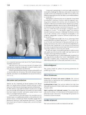
final periapical radiograph was obtained. The authors thank the patient for granting permission for
Clinical and radiographic follow-up were performed 6 and publishing this case.
12 months after the procedures. The tooth remained asymp-
tomatic, and a final coronal rehabilitation was performed (Fig-
ures 4 e 5). Ethical disclosures
Protection of human and animal subjects. The authors
Discussion and conclusions declare that no experiments were performed on humans or
animals for this study
Studies on the complexity of dental anatomy have shown
Confidentiality of data. The authors declare that they have
that root canals present different internal anatomical varia- followed the protocols of their work center on the publication
tions due to the natural physiological process of formation. of patient data.
1,2
1
In 1984, Vertucci reported that incisors usually have a single
root and a single canal. However, the presence of extra canals Right to privacy and informed consent. The authors have
7
6
has been found in double and triple central incisors, and obtained the written informed consent of the patients or sub-
even in quadruple lateral incisors. 8 jects mentioned in the article. The corresponding author is in
These variations in the root canals of the maxillary central possession of this document.
incisors may occur unilaterally, similar to the one found in the
4
present report, or bilaterally. These variations apply both to
10
9
permanent dentition and deciduous dentition. The extra Conflict of interest
root canals can be found simultaneously in central and later-
al incisors. 11 The authors have no conflicts of interest to declare.