


Page 41 - SPEMD_62-3
P. 41
rev port estomatol med dent cir maxilofac . 2021;62(3):163-169         167


                                                               thin radiopaque halo and no root resorption or displacement
                                                               of the associated dental elements 13 and 15, despite bulging
                                                               of the cortical bone. The association with dental apices and
                                                               the indolent clinical -radiographic behavior of the COC led to
                                                               the clinical hypothesis of a radicular cyst.
                                                                 The literature recommends enucleation associated with
                                                               lesion curettage, although marsupialization/decompression is
                                                               also among the treatments of choice for larger lesions to reduce
                                                               cyst size and surgery length. This maneuver has improved
                                                                                                             22
                                                               prognosis, leading to low recurrence rates (approximately 5%).
                                                               In this report, due to the lesion’s size and well -circumscription,
                                                               enucleation associated with curettage was performed. The suc-
                                                               cess of this conservative treatment is evidenced by the lack of
                                                               recurrences in the last 3 years of follow -up.
                                                                 COCs may be associated with other odontogenic lesions,
                                                               and the literature commonly reports association with odonto-
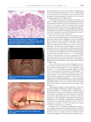
            Figure 10. Antral pseudocyst. Details of the non-  mas and ameloblastomas. Dentigerous cysts are considered
            -keratinized stratified squamous epithelium of the antral   the most frequent cysts. In addition, impacted or dislocated
            pseudocyst. (Hematoxylin -eosin; scale barr: 200µm in A,   adjacent teeth are also frequent.  In another study, three
                                                                                          23
            100µm in B, 500µm in C, and 100µm in D).
                                                               COCs were associated with odontomas and one with amelo-
                                                                       24
                                                               blastomas.  The present case report suggests an association
                                                               between a COC and an AP, which has not been mentioned in
                                                               the literature. APs are solitary, dome -shaped radiopaque mass-
                                                               es located on the maxillary sinus floor that can further reduce
                                                               the size of this anatomical structure. 11-13,25-27  This finding is
                                                               consistent with the present case since a veiling of the maxil-
                                                               lary sinus caused by the AP was observed, and the central ra-
                                                               diopaque area in the maxillary sinus led us to hypothesize a
                                                               diagnosis of COC for the lesion in the antrum (rather than the
                                                               periapical one).
                                                                 APs are often detected on routine radiographic examina-
                                                               tions. Surgical excision in these cases is unnecessary, as stud-
                                                               ies have shown no  progression or even regression  during
                                                               follow -up. 11,12,25  When present, symptoms range from head-
                                                               aches to face pain and nasal obstruction and discharge. 12,25  In
                                                               some cases, a positive aspiration mimicking a cystic lesion
                                                                           13
                                                               may be present.  Given these clinical and radiographic find-
            Figure 11. Extraoral clinical image without facial   ings, the literature indicates mucocele of the sinus and odon-
            asymmetry.                                         togenic cysts as differential diagnoses. 11,13  Due to the mixed
                                                               radiographic aspect of the AP in the maxillary sinus region, the
                                                               clinical hypothesis of COC was raised. The literature reports
                                                               that COCs can present at this location and even mimic a sinus
                                                               mucocele. 3,28
                                                                 Although APs’ etiology is not well understood, other stud-
                                                               ies report allergies, barotrauma, rhinitis, rhinosinusitis, 12,25
                                                               periapical infections, 10,11,25  periodontitis, 10,25  viral infections of
                                                               the respiratory tract, and mucosal irritations caused by lack of
                                                               air humidity  as possible factors. Since COCs are a noninflam-
                                                                        13
                                                               matory odontogenic lesion, they are unlikely to cause APs’
                                                               development. The radiographic appearance observed in the
                                                               present case, which indicates a clear separation between the
                                                               two lesions, may support this hypothesis. Further medical in-
                                                               vestigation is required to confirm if the patient has a history
                                                               of allergies or respiratory tract infections.
                                                                              29
                                                                 Beaumont et al.  recommend a Caldwell -Luc surgery or
                                                               sinus endoscopic surgery as the ideal treatment in cases of
                                                               sinus graft when APs are present and advocate the complete
            Figure 12. Intraoral image after 5 years of follow -up   removal of the sinus lining to prevent a recurrence. These
            post -surgery.
                                                               authors also suggested at least 6 months of healing after le-

