


Page 40 - SPEMD_62-3
P. 40
166                    rev port estomatol med dent cir maxilofac. 2021;62(3):163-169


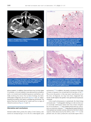
            Figure 6. Postoperative computerized tomography    Figure 8. Calcifying odontogenic cyst. Numerous ghost
            without maxillary sinus veiling.                   cells within the epithelial component (black arrows),
                                                               organized as fused cell masses forming large sheets of
                                                               amorphous and acellular material in the fibrous
                                                               connective tissue capsule and calcifications (red arrows).


            Figure 7. Calcifying odontogenic cyst. Pathological cavity   Figure 9. Antral pseudocyst. Multiple epithelium -lined
            lined with ameloblastomatous basal layer epithelium   cystic compartments and loose connective tissue (blue
            (stars). Overlying layer cells are loosely organized,   stars) presenting mononuclear inflammatory infiltrates
            resembling the stellate reticulum.                 (green stars) and extravasation of mucin -compatible
                                                               eosinophilic material (yellow stars).


           phous material. In addition, the maxillary sinus had two types   and tumors. 15,16  In addition, the peak occurrence of this type
           of epithelia: a ciliated cylindrical pseudostratified epithelium   of lesion is between the second and third decades of life. 8,17
           and a non -keratinized stratified squamous epithelium, par-  Thus, since the patient in this case report was 66 years old at
           tially involving the lesion. Due to these findings, the histo-  the time of diagnosis, he may have been diagnosed late,
           pathological diagnosis was an AP (Figures 9 and 10). Subse-  which would explain the greater bone growth and expansion
           quently, enucleation with lesion curettage was performed. The   observed.
           patient has been followed up for 5 years and has no signs of   COCs may be intraosseous or peripheral, the latter being
           clinical recurrence (Figures 11 and 12).           extremely rare. 1,17  Radiographic findings include multilocular
                                                              or unilocular radiolucent lesions that may contain irregular
                                                              foci of calcifications. 1-3,18  Teeth displacement and root resorp-
           Discussion and conclusions                         tion are relatively common. 19,20  Regarding biological behavior,
                                                              COCs present an indolent course, contrary to the DGCT, which
           Several studies have proven the rarity of COC lesions, esti-  explains their distinction by the WHO in 2017. 1-2,6,8,21  In the
           mated as corresponding to 1% to 3% of all odontogenic cysts   present case, the lesion presented a unilocular aspect with a

