


Page 39 - SPEMD_62-3
P. 39
rev port estomatol med dent cir maxilofac . 2021;62(3):163-169         165


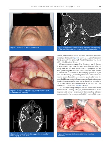
            Figure 1. Swelling in the right hemiface.           Figure 4. Hypodense lesion causing maxillary sinus veiling
                                                                in the sagittal section of the computerized tomography.


                                                               formed, and the entire lesion was sent for further anatomo-
                                                               pathological analysis (Figures 5 and 6). In addition, the canine
                                                               dental element was extracted. Finally, the suture was made
                                                               with a 3 -0 silk suture thread.
                                                                 Light microscopy analysis of the first lesion revealed a cys-
                                                               tic lesion of odontogenic origin characterized by a pathological
                                                               cavity lined with both a stratified and a simple paved epithe-
                                                               lium, presenting cells from the columnar basal layer, similar
                                                               to ameloblasts. In some areas, the overlying epithelium layers
                                                               were loosely arranged, resembling the stellate reticulum of the
                                                               enamel organ. In addition, numerous ghost cells were ob-
                                                               served within the epithelial component as fused cell masses,
                                                               forming large sheets of amorphous and acellular material in
                                                               the fibrous connective tissue capsule. These findings con-
                                                               firmed the COC diagnosis (Figures 7 and 8).
                                                                 The histopathology analysis of the associated lesion
                                                               demonstrated a loosely arranged, swollen connective tissue
            Figure 2. Increased intraosseous growth volume and
            normochromic staining.                             fragment with intense mononuclear inflammatory infiltrate
                                                               areas permeating large amounts of slightly eosinophilic amor-


            Figure 3. Presence of material suggestive of maxillary   Figure 5. Trans -surgical enucleation and curettage
            sinus injury in axial cuts.                         procedure.

