
Page 59 - SPEMD_62-1
P. 59
rev port estomatol med dent cir maxilofac . 2021;62(1):50-55 53
blood examination showed nothing abnormal. Based on clin-
ical and radiologic aspects, the initial diagnosis was keratocyst
or ameloblastoma, and surgical treatment was indicated after
establishing the final diagnosis.
An incisional biopsy was performed under local anesthe-
sia. After the mucoperiosteal flap was raised, a discharge of a
whitish pasty material, suggestive of keratin, was observed.
The anatomopathological examination confirmed the initial
diagnosis of keratocyst (Figure 7).
The patient was informed about all treatment modalities and
the recurrence rates associated with each one. Although partial
resection with the placement of a customized prosthesis was Figure 9. 18 -month panoramic radiograph follow -up
recommended, he opted for a more conservative treatment, with showing significative bone neoformation with no signs of
recurrence.
enucleation followed by the application of Carnoy’s solution.
An incision was made, under general anesthesia, on the
anterior border of the ramus, extending to the second premo-
lar area. The mucoperiosteal flap was raised, followed by com-
plete enucleation of the lesion. Then, the bone cavity was coat-
ed with gauze, and the Carnoy’s solution was applied for 3
minutes, followed by saline irrigation; this procedure was re-
peated three times. The wound was closed with absorbable
suture, and the healing was uneventful. The patient was dis-
charged from the hospital 24 hours later, with no pain com-
plaints but mentioning lower lip cushioning.
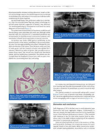
Figure 10. A: sagittal section of the initial tomography
showing extensive bone destruction; B: sagittal section of
the same area, 18 months later, showing almost
complete bone neoformation and no signs of recurrence.
In a seven -day postoperative examination, he had good
local healing and reported improvement of the cushioning.
Complete resolution of paresthesia occurred around 40 days
after surgery.
The patient remains in semiannual radiographic control
(Figures 8 and 9) and 18 -month cone -beam tomography con-
Figure 7. Thin cystic lesion lining epithelium with a
hyperchromatic basal layer in palisade and corrugated trol. He shows significant bone neoformation compared to the
parakeratin layer. initial tomography, with no signs of recurrence (Figure 10).
Discussion and conclusions
Early diagnosis of OK is the best way to avoid extensive bone
destruction and more aggressive surgery. Unfortunately, in
this case, despite the large radiolucent area in the mandibular
ramus, three different professionals missed it. This overlook
calls attention to the fact that some dentists look for alter-
ations only in the teeth without analyzing all the anatomic
structures in a panoramic radiograph.
Many accepted methods are used in the treatment of OKs,
and the greatest challenge is to completely remove the cystic
Figure 8. 6 -month panoramic radiograph follow -up capsule, which is thin and friable. More conservative modal-
showing bone neoformation.
ities include marsupialization, enucleation with or without