
Page 37 - SPEMD_62-1
P. 37
rev port estomatol med dent cir maxilofac . 2021;62(1):29-34 31
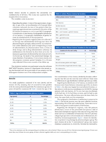
tients’ clinical records to preserve the anonymity and Table 2. Lichen Planus lesions by location.
confidentiality of all data. This study was approved by the
Hospital Ethics Committee. Lichen planus lesions’ location N Percentage
The variables under study were:
Skin 132 75.9%
– Regarding the patient: 1) date of the appointment; 2) gen- Oral mucosa 4 2.3%
der; 3) age of the 1st manifestation of LP (except when Genital mucosa 4 2.3%
otherwise indicated, the date of the patient’s first Der-
matology appointment was considered); 4) location of the Skin + oral mucosa 20 11.5%
LP’s lesions (cutaneous vs. oral vs. genital); 5) topograph- Skin + oral mucosa + genital mucosa 5 2.9%
ic distribution of skin lesions; 6) topographic distribution
of oral lesions; 7) topographic distribution of genital le- Skin + genital mucosa 8 4.6%
sions; 8) administration of therapy (yes/no). Oral + genital mucosa 1 0.6%
– Regarding the therapeutic regimen (data not collected if
no therapy was administered): 9) therapy start date; 10) TOTAL 174 100.0%
pharmacological group(s) administered; 11) corticoste-
roid active substance (AS)* (and corresponding a) route
of administration; b) pharmaceutical form; c) dose; d) Table 3. Lichen Planus lesions’ location in the oral cavity.
daily dosage; and e) drug administration scheme); 12) Locations in the oral cavity N Percentage
antihistamine AS (followed by data from a) to e)*); 13)
immunosuppressant AS (followed by data from a) to e)*); Buccal mucosa 21 70.0%
14) follow-up; 15) result of the established therapy; 16) Palate 1 3.3%
mean duration of symptoms; 17) recurrence events; and
18) symptom remission period. Variables 15 to 18 were Tongue 2 6.7%
only collected if there were records of the follow -up. Buccal mucosa, palate and tongue 1 3.3%
The statistical analysis was performed using the software Buccal mucosa, tongue and alveolar ridge 1 3.3%
IBM SPSS Statistics, version 25. Hypotheses were tested us- No information 4 13.3%
®
®
ing the binomial and chi -square tests of one sample and the
Kolmogorov-Smirnov test of two independent samples. TOTAL 30 100.0%
first manifestation of the disease, divided by decades, reflect-
Results ing that the 4th and 5th were the most affected. There were
no differences in age distribution between genders (p=0.989).
The study population consisted of 85 men (48.9%) and 89 Regarding the distribution of LP lesions, no statistical dif-
women (51.1%), meaning that LP occurred in both genders ferences were found between trimesters (p=0.235). As shown
with the same probability (p=0.820). The minimum age was 11 in Table 2, the skin was largely the most affected location, ei-
years old and the maximum 88. Table 1 shows the age of the ther alone or with mucous membranes (n=165;94.9%). Among
patients with skin lesions, 40.7% only had one site affected,
Table 1. Age of onset of lichen planus, in years. 22.8% had two, 32.5% had three or more, and 4% had no site
information. The most affected sites were the lower limb, the
Age (years) N Percentage
torso, the upper limb, and the extremities.
11-20 8 4.6% The topographic distribution of oral lesions is presented in
Table 3. The buccal mucosa was the most affected location,
21-30 22 12.6%
representing 70% of the oral cavity lesions. There were no re-
31-40 25 14.4% cords of malignant transformation of OLP lesions.
Regarding the therapeutic regimen, only 166 patients
41-50 40 23.0%
(95.4%) had a record of the prescribed therapeutics. Half of
51-60 39 22.4% them had a therapeutic regimen with only one drug, while the
other half had a combination of medicines. The main pharma-
61-70 28 16.1%
cological groups were corticosteroids and antihistamines, fol-
71-80 10 5.7% lowed by immunosuppressants. Figure 1 shows the distribu-
80+ 2 1.1% tion of patients’ drug therapy by pharmacological group. The
most chosen AS in the “1 corticosteroid” group was topical
TOTAL 174 100.0%
clobetasol propionate (n=30;38.0%), followed by betametha-
* The active substance and the corresponding a) to e) data were only collected if there was any AS from that pharmacological group. If there were
more than one AS from the same group, all were collected together with each one’s corresponding a) to e) data.