
Page 38 - SPEMD_62-1
P. 38
32 rev port estomatol med dent cir maxilofac. 2021;62(1):29-34
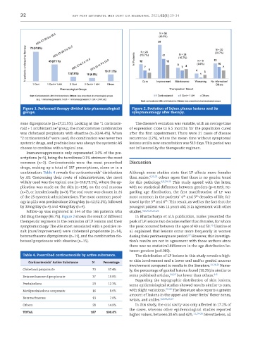
Figure 1. Performed therapy divided into pharmacological Figure 2. Evolution of lichen planus lesions and its
groups. symptomatology after therapy.
sone dipropionate (n=17;21.5%). Looking at the “1 corticoste- The disease’s evolution was variable, with an average time
roid + 1 antihistamine” group, the most common combination of expression close to 6.5 months for the population cured
was clobetasol propionate with ebastine (n=20;44.4%). When after the first appointment. There were 21 cases of disease
“2 corticosteroids” were used, the combination was never two recurrence (12%), where the mean time without symptoms/
systemic drugs, and prednisolone was always the systemic AS lesions until a new exacerbation was 513 days. This period was
chosen to combine with a topical one. not influenced by the therapeutic regimen.
Immunosuppressants only represented 3.0% of the pre-
scriptions (n=5), being the tacrolimus 0.1% ointment the most
common (n=3). Corticosteroids were the most prescribed Discussion
drugs, making up a total of 187 prescriptions, alone or in a
combination. Table 4 reveals the corticosteroids’ distribution Although some studies state that LP affects more females
by AS. Concerning their route of administration, the most than males, 4,20,23 others agree that there is no gender trend
widely used was the topical one (n=148;79.1%), where the ap- for this pathology. 6,8,24-26 This study agreed with the latter,
plication was made on the skin (n=138), on the oral mucosa with no statistical difference between genders (p=0.820). Re-
(n=7), or intralesionally (n=3). The oral route was chosen in 24 garding age distribution, the first manifestation of LP was
th
th
of the 25 systemic administrations. The most common posol- most common in the patients’ 4 and 5 decades of life, fol-
th
ogy in pills was prednisolone 20mg/day (n=12;52.2%), followed lowed by the 3 and 6 . This result, as well as the fact that the
rd
by 30mg/day (n=4) and 40mg/day (n=4). youngest patient was 11 years old, is in agreement with other
Follow -up was registered in 144 of the 166 patients who studies. 4,8,20,23,25,26
did drug therapy (86.7%). Figure 2 shows the result of different In Bhattacharya et al.’s publication, males presented the
therapeutic regimens in the remission of LP lesions and their peak of LP lesions two decades earlier than females, for whom
25
symptomatology. The ASs most associated with a positive re- the peak occurred between the ages of 40 and 50. Usatine et
sult (cure/improvement) were clobetasol proprionate (n=14), al. explained that lesions occur more frequently in women
27
betamethasone dipropionate (n=11), and the combination clo- during their perimenopause period. However, this investiga-
betasol proprionate with ebastine (n=15). tion’s results are not in agreement with those authors since
there was no statistical difference in the age distribution be-
tween genders (p=0.989).
Table 4. Prescribed corticosteroids by active substance. The distribution of LP lesions in this study reveals a high-
Corticosteroids’ Active Substance N Percentage er skin involvement and a lower oral and/or genital mucous
involvement compared to results in the literature. 11,24,25 Name-
Clobetasol propionate 70 37.4% ly, the percentage of genital lesions found (10.3%) is similar to
Betamethasone dipropionate 37 19.8% some published articles, 24,25 but lower than others. 4-6
Regarding the topographic distribution of skin lesions,
Prednisolone 23 12.3%
some epidemiological studies showed results similar to ours,
Metilprednisolone aceponate 16 8.6% with slight variations. 24-26 The literature also reports a greater
amount of lesions in the upper and lower limbs’ flexor zones,
Betamethasone 13 7.0% 6,8,24,25,27
wrists, and ankles.
Others 28 14.9% In this study, the oral cavity was only affected in 17.2% of
the cases, whereas other epidemiological studies reported
TOTAL 187 100.0% 11,24-26
higher values, between 20.6% and 42%. Nevertheless, all