
Page 60 - SPEMD_60-4
P. 60
208 rev port estomatol med dent cir maxilofac. 2019;60(4):205-209
Discussion and conclusions
The cells that originate trichilemmoma appear to be locat-
ed in a superficial area of the hair follicle, just below the
basement membrane, at the level of the sebaceous gland.
There is a lobular proliferation of small uniform cells to the
dermis from the epidermis, and the presence of clear cells
containing glycogen is the most notable alteration. 1-4
The relationship between trichilemmoma and common
wart is controversial, and trichilemmomas are actually often
clinically diagnosed as warts. On the other hand, areas his-
tologically similar to trichilemmomas are often found in warts.
Some authors, therefore, believe that trichilemmomas may
correspond to mere mature warts, with some period of evolu-
9
tion, that have suffered trichilemmal differentiation. Howev-
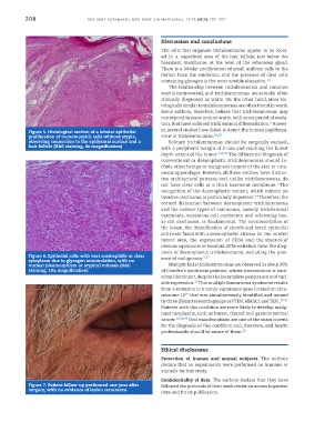
Figure 5. Histological section of a lobular epithelial er, several studies have failed to detect the human papilloma-
proliferation of monomorphic cells without atypia, virus in trichilemmomas. 28,29
observing connection to the epidermal surface and a Solitary trichilemmomas should be surgically excised,
hair follicle (H&E staining, 4x magnification) with a peripheral margin of 2 mm and reaching the fullest
depth extent of the tumor. 4,30,31 The differential diagnosis of
conventional or desmoplastic trichilemmomas should in-
clude other benign or malignant tumors of the skin or cuta-
neous appendages. However, all these entities have distinc-
tive architectural patterns and, unlike trichilemmomas, do
3
not have clear cells or a thick basement membrane. The
recognition of the desmoplastic variant, which mimics an
14
invasive carcinoma, is particularly important. Therefore, the
correct distinction between desmoplastic trichilemmoma
and the various types of carcinoma, namely trichilemmal
carcinoma, squamous-cell carcinoma and sclerosing bas-
al-cell carcinoma, is fundamental. The circumscription of
the lesion, the identification of chords and small epithelial
cell nests fused with a desmoplastic stroma in the central
tumor area, the expression of CD34 and the absence of
obvious squamous or basaloid differentiation favor the diag-
nosis of desmoplastic trichilemmoma, excluding the pres-
Figure 6. Epithelial cells with vast eosinophilic or clear 3,32
cytoplasm due to glycogen accumulation, with no ence of malignancy.
nuclear pleomorphism or atypical mitoses (H&E Multiple facial trichilemmomas are observed in about 99%
staining, 10x magnification) of Cowden’s syndrome patients, whose transmission is auto-
somal dominant, despite the incomplete penetrance and vari-
33
able expression. This multiple hamartoma syndrome results
from a mutation in a tumor suppressor gene located on chro-
34
mosome 10 that was simultaneously identified and named
by three distinct research groups as PTEN, MMAC1 and TEP1. 35-37
Patients with this condition are more likely to develop malig-
nant neoplasms, such as breast, thyroid and gastrointestinal
cancer. 10,38,39 Oral manifestations are one of the main criteria
for the diagnosis of this condition and, therefore, oral health
professionals should be aware of them. 10
Ethical disclosures
Protection of human and animal subjects. The authors
declare that no experiments were performed on humans or
animals for this study.
Confidentiality of data. The authors declare that they have
Figure 7. Patient follow-up performed one year after followed the protocols of their work center on access to patient
surgery, with no evidence of lesion recurrence
data and for its publication.