
Page 33 - SPEMD_58-4
P. 33
rev port estomatol med dent cir maxilofac . 2017;58(4):219-224 221
stored in a glass receptacle in a 10% formaldehyde solution for Table 1. Experimental groups according to the accessory
seven days and kept refrigerated. used, composition, fabrication and manufacturing lot.
The minimum sample size required for this study was cal-
culated using the following parameters: a test power of 80% (β Groups Accessories Composition Brand Lot
= 0.20) and an error of 5% (α=0.05). The calculation determined Used
a minimum sample size of 12 samples. Considering the possi- 1 Lingual button Polycarbonate Morelli 1801732
bility of using non-parametric statistics (Friedman test), an in- Composite with glass fiber
crease of 20% was applied, which resulted in the adjustment of Hook for
the minimum size required for 15 individuals. The sample cal- 2 traction Stainless Steel Morelli 1767974
culation was performed in G*Power (version 3.1.9.2, Germany). in impacted
The teeth were embedded in PVC rings (Tigre, Joinville, Bra- teeth
zil) with acrylic resin (Clássico, São Paulo, Brazil) so that only 3 Hook with chain Stainless Steel Morelli 1809417
their crowns were exposed. In order to maintain the surface
to be glued to the bracket perpendicular to the ground, a 4 Cleat Stainless Steel Morelli 1827790
square was used for standardization. After polymerizing the 5 Brackets Stainless Steel Morelli 1828477
resin, all the test specimens were stored in distilled water and Convex lingual
again placed in the refrigerator at 5ºC. 6 button Stainless Steel Morelli 1809815
The vestibular surfaces of the teeth received prophylaxis
with a rubber cup (Viking, KG Sorensen, Barueri, Brazil), an 7 Concave lingual Stainless Steel Morelli 1834886
button
extra-fine pumice stone (S.S.White, Juiz de Fora, Brazil) and
distilled water for 15 seconds. Afterwards, they were washed 8 Mesh Plaited wires Morelli 1430356
with air spray/distilled water and dried with oil- and humidi- Stainless Steel
ty-free jets of air for the same length of time. After prophylax-
is, etching was performed with 37% phosphoric acid (FGM,
Joinville, Brazil) for 30 seconds, followed by washing with wa- the mesh was placed on the tooth surface coupled with an
ter and drying with jets of air for the same period. A thin coat elastic, and the interior of the elastic was filled with orthodon-
of primer (Transbond XT; 3M Unitek, Monrovia, California, tic composite (Transbond XT, Monrovia, USA).
USA) was applied and light polymerized for 40 seconds. The next step was light polymerization for 40 seconds with
Before the bonding process, the teeth were randomly di- the light-polymerization device XL 1500 (3M Unitek, Monrovia,
vided into groups corresponding to the accessory that would CA, USA), using a light intensity of 400 mW/cm², regularly
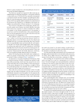
be bonded to them, as shown in Table 1 and Figure 1. checked with a radiometer (Demetron, Danbury, CT, USA).
An orthodontic composite (Transbond XT, Monrovia, USA) After bonding, the test specimens were stored in artificial
was used for bonding. Initially, composite was applied on the saliva and kept in an oven at a temperature of 37°C for 24
base of the accessories, which were then placed on the tooth hours. The bond strength tests were performed in a universal
surface and subjected to a force of 300g, to allow extravasation mechanical test machine (AME-2kN; Fillizola, São Paulo, Bra-
of the excess composite. For this purpose, a weight of 300 g zil), operating at a speed of 0.5 mm/min, by means of a chis-
was positioned on top of the brackets. The excess composite el-shaped active tip. The shear strength forces were obtained
was removed with an exploratory probe no. 5. in kilogram-force, converted into Newton, and divided by the
The mesh was previously standardized so that it would base area of the tested accessory. Thus, the results were given
always have the same dimensions, and the same amount of in megapascal (MPa). Megapascal was the chosen unit because
orthodontic composite would be applied. For this purpose, the it allowed individualizing the force applied on a specific area
internal diameter of an orthodontic elastic (3.1 mm) was used in mm², thereby annulling the variable corresponding to the
to delimit the cross-sectional area of the mesh. Afterwards, base area of each accessory tested.
After performing the shear strength tests, the vestibular
surfaces of the tested specimens were evaluated under a ste-
reomicroscope (Carl Zeiss, Göttingen, Germany), at 16x mag-
nification, to quantify the ARI. The ARI scores ranged from 0
to 3, with 0 indicating that there were no composite remnants
on the enamel, 1 that there was less than half of the compos-
ite, 2 that there was more than half of the composite and 3 that
the whole composite was on the tooth surface.
The means and standard deviations were calculated for the
descriptive analysis of the shear strength and ARI. For the in-
ferential analysis of the shear strength, the homogeneity of
variances was tested using the Levene’s test and the normality
Figure 1. Accessories used: 1 – composite lingual button; of the residues using the analysis of variance (one-way ANO-
2 – hook for applying traction to impacted teeth; 3 – hook VA), namely, the Kolmogorov-Smirnov test. After verifying the
with chain; 4 – cleat; 5 – bracket; 6 – convex lingual heterogeneity of variances and the asymmetrical distribution
button; 7 – concave lingual button; 8 – mesh. of the residues, the Kruskal-Wallis test was applied to compare