Page 71 - SPEMD_58-2
P. 71
rev port estomatol med dent cir maxilofac . 2017;58(2):126-131 127
sional, o tumor foi diagnosticado como TOA. A enucleação da lesão e do canino impactado
foi realizada. Quatro meses depois, o paciente não apresentou sinais de recorrência e foi
encaminhado para tratamento ortodôntico. Um novo procedimento cirúrgico foi realizado
para remover a gengiva que cobria os pré-molares para promover sua erupção. Após cinco
anos, os pré-molares encontram-se em posição no arco dentário e não existem sinais de
recorrência da lesão. (Rev Port Estomatol Med Dent Cir Maxilofac. 2017;58(2):126-131)
© 2017 Sociedade Portuguesa de Estomatologia e Medicina Dentária.
Publicado por SPEMD. Este é um artigo Open Access sob uma licença CC BY-NC-ND
(http://creativecommons.org/licenses/by-nc-nd/4.0/).
palpation. The buccal cortical plate of the right maxilla was
Introduction expanded from the central incisor to the first molar and palatal
Odontogenic tumors and hamartomas are included in a wide displacement of lateral incisor on the same side (Figure 1). Fa-
variety of rare lesions that originate from odontogenic tissues cial asymmetry was not observed upon extra oral examination.
and are present at variables levels of differentiation. The de- The panoramic radiography showed a well -defined, uni-
termination of its exact nature (i.e., hamartoma or neoplasm) locular radiolucency in the maxilla associated with the
is diffcult and often inconclusive, which makes the nomen- unerupted right upper canine, with no evidence of calcifica-
clature for these types of lesions diffcult. 1 tions or root resorptions. It also showed the displacement of
The Adenomatoid odontogenic tumor (AOT) was first report- first and second premolars on the same side and a delay in its
2
ed in 1907, called the pseudo -adenoameloblastoma. Different eruption process (Figure 2). Radiographic and clinical findings
nomeclatures have also been used to describe this tumor: ade- were compatible with the diagnosis of dentigerous cyst, AOT,
noameloblastic and ameloblastic adenomatoid tumor. The term and unicystic ameloblastoma.
Adenomatoid odontogenic tumor, adopted by the World Health Fine needle aspiration was negative and an incisional bi-
Organization classification of odontogenic tumors in 1971, is still opsy was performed. Histological examination revealed cuboi-
the most accepted and currently used nomenclature. 3,4 dal or spindle -shaped epithelial cells forming aggregates or
AOT is a rare and non -aggressive epithelial odontogenic typical rosette -like structures with minimal connective tissue,
tumor that occurs in intraosseous and peripheral forms. The and cuboidal or low columnar cells forming glandular duct-
intraosseous variants are the most frequent and include fol- -like structures, confirming the diagnosis of an AOT (Figure 3).
licular and extrafollicular types. Radiographically, the central After confirming the diagnosis, the enucleation of the lesion
lesions (intraosseous) appear as a unilocular and radiolucent was performed under local anesthesia, together with the re-
area, which is well -defined and often associated with an im- moval of the impacted canine and deciduous molars (Figure 4).
pacted tooth. Two -thirds of intraosseous cases present radi- The final diagnosis of an intraosseus follicular variant of AOT
opacity within them. 5 was reconfirmed after the specimen had been microscopically
This type of lesion corresponds to 2% to 7% of all odonto- examined.
genic tumors, usually affecting young patients, mostly during
their second and third decades of life. Women are affected
more often than men (a ratio of 1.9:1.0), and the lesions tend
to occur in the anterior maxilla region. 4,6
The aim of this study was to report the surgical manage-
ment of an AOT, show the results from the five years of follow-
-up, and discuss clinical and radiographic aspects with the
current literature.
Case report
An 11 -year -old female patient was referred to the Oral and
Maxillofacial Surgery service of the School of Dentistry of the
Federal University of Minas Gerais (UFMG), Belo Horizonte,
Brazil, presenting a 5 -6 -month history of asymptomatic tu-
mor growth on the right side of the maxilla. There was no
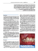
history of trauma and the lesion had progressively increased Figure 1. Clinical image showing right anterior
in size in recent months. maxillary swelling with the expansion of the buccal
Clinical examination revealed swelling with ill -defined cortical plate, palatal displacement of the lateral incisor,
margins, with normal overlying mucosa, which was firm on and the absence of upper right canines and premolars.