Page 72 - SPEMD_58-2
P. 72
128 rev port estomatol med dent cir maxilofac. 2017;58(2):126-131
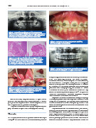
Figure 2. Panoramic radiography shows the unilocular
lesion surrounding the coronal impacted right upper canine
beyond the amelocemental junction and the displacement
of the first and second premolars on the same side.
Figure 5. Five year clinical postoperative follow up
showed no signs of recurrence.
Figure 3. A: Higher power (x100) histology shows a
hematoxylin and eosin stained section with a closer
look at the spindle shaped epithelial cells arranged
in whorls and strands. B: A duct like structure lined
by one layer of cuboidal epithelial cells.
Figure 6. Five year postoperative panoramic radiography
shows normal healing and no signs of recurrence.
histopathological characteristics and similarity to ameloblas-
toma, thus previously receiving the name of pseudo-
-adenomeloblastoma. When compared to ameloblastomas,
2
the most common odontogenic tumor, AOT is a nonaggres-
sive tumor, encapsulated with limited growth and no tenden-
cy of recurrence. It is usually associated with an unerupted
Figure 4. A: Enucleation of the lesion and associated permanent tooth. Radiographically, in most cases, AOT shows
impacted canine. B: Gross examination of the specimen a unilocular radiolucency with well -defned borders and may
showed a single, well encapsulated soft tissue. C: 4,7
Immediate postoperative photography. contain numerous dispersed radiopaque foci. Except for the
absence of calcifcations in the lesion, the present case pre-
sents the classic characteristics of the lesion.
Neoplastic or hamartous lesions can develop at any stage
8
Four months later, the patient showed no signs of recur- of a complex process called odontogenesis. In relation to the
rence and was referred for orthodontic treatment. A new sur- theory about the origin and pathogenesis of AOT, it seems that
gical procedure was performed to remove the gums that cov- this tumor is derived from the odontogenic epithelium of the
ered the premolars and promote their eruption. dental lamina complex or its cellular remnants situated in the
The patient underwent follow -up and, five years after sur- gubernacular cord. 9
gery, has shown no signs of recurrence (Figures 5 and 6). The gubernacular cord is a fibrous innerved, vascularized,
and lymphatic channel with epithelial cells or cell clusters
from the fragmented dental lamina running in a bony channel
Discussion called the gubernacular canal, which connects the pericoronal
follicular tissue of the permanent tooth to the alveolar crest
AOT is a rare, hamartomatous, epithelial lesion of odontogen- and the palatal gingiva of the deciduous tooth. Some authors
4,6
ic origin. Its one of the most controversial lesions, due to its conclude that dental lamina in the gubernacular cord of the